
Abstract
Chronic wasting disease (CWD) is the only known transmissible spongiform encephalopathy affecting free-ranging wildlife. Although the exact mode of natural transmission remains unknown, substantial evidence suggests that prions can persist in the environment, implicating components thereof as potential prion reservoirs and transmission vehicles. CWD-positive animals may contribute to environmental prion load via decomposing carcasses and biological materials including saliva, blood, urine and feces. Sensitivity limitations of conventional assays hamper evaluation of environmental prion loads in soil and water. Here we show the ability of serial protein misfolding cyclic amplification (sPMCA) to amplify a 1.3 x 10⁻⁷ dilution of CWD-infected brain homogenate spiked into water samples, equivalent to approximately 5 x 10⁷ protease resistant cervid prion protein (PrPCWD) monomers. We also detected PrPCWD in one of two environmental water samples from a CWD endemic area collected at a time of increased water runoff from melting winter snow pack, as well as in water samples obtained concurrently from the flocculation stage of water processing by the municipal water treatment facility. Bioassays indicated that the PrPCWD detected was below infectious levels. These data demonstrate detection of very low levels of PrPCWD in the environment by sPMCA and suggest persistence and accumulation of prions in the environment that may promote CWD transmission.
Acknowledgements
The authors thank the North American Deer Farmers Association, the Colorado Section of the American Water Resources Association, the City of Fort Collins Water Board and the Colorado State University College Research Council and Infectious Disease Initiative. We thank Tom Holley and Les Nichols for help with water collections, Kathleen Ganzer, Judy Billica and Grant Jones for help with water quality analyses and Terry Spraker for CWD-infected brain samples, technical assistance and advice.
Figures and Tables
Figure 1 Normal brain homogenate negative controls. All samples were digested with Proteinase K except normal brain homogenate (NBH) in lane 1. NBH, Normal brain homogenate control. +, 1:100,000 positive amplification control. Molecular weight markers in kilodaltons are shown to the left of the blots, which show thirty samples representative of 241 NBH negative controls. All samples were negative after six rounds of sPMCA except the sample in Gel C, lane 6.
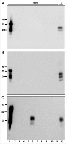
Figure 2 PrPCWD detection limit in water. (A) All samples were digested with Proteinase K except normal brain homogenate (NBH) in lane 1. Lanes 2 through 12 show amplified samples at the indicated starting dilution of CWD-positive brain into water. Lanes 13 and 14 show amplified NBH controls. The highest dilution reproducibly detected was 1:1.3 × 10⁷ (lane 9). (B) Representative western blot showing serial 2-fold dilutions of PrPCWD (lanes 1–6) and rCerPrP (lanes 8–12). Samples were PK digested where indicated. Known quantities of rCerPrP were loaded in the indicated lanes to generate the standard curve in shown in (C). Densitometric analyses were performed on four unsaturated replicate blots. PRPCWD was estimated by interpolation of band intensities to the standard curve. The linear regression equation, the standard deviation of the residuals (Sy.x) and the goodness-of-fit (r2) of the curve are indicated. Molecular weight markers in kilodaltons are shown to the left of the blots, which are representative of four replicates using two distinct CWD prion isolates.
Figure 3 PrPCWD amplification in raw water samples from non-CWD-endemic areas. All samples were digested with Proteinase K except normal brain homogenate (NBH) in lane 1. Lane 2 shows amplified NBH control. sPMCA failed to amplify any PrPCWD from five replicate samples (lanes 3 to 7) of raw water from areas in IL (A, ground water and B, surface water) and Michigan (C, Lake Michigan and D, surface water) without any reported CWD cases, but amplified dilutions of PrPCWD spiked into these samples (log10, lanes 8 to 12) with similar efficiency as PrPCWD spiked into purified water. Data are representative of four separate experiments.
Figure 4 Locations of the Cache La Poudre river, Horsetooth reservoir and the Fort Collins Water Treatment Facility in the South Platte river basin watershed. (A) Map showing the South Platte river basin watershed. Cache La Poudre river (PR) and Horsetooth reservoir (HT) are shown 2X scale for clarity. The Colorado-Big Thompson western slope watershed, which feeds HT, is shown in dark blue (top left watershed on the map). The Cache la Poudre watershed is shown in purple (top right watershed). FC, city of Fort Collins; FCWTF, Fort Collins water treatment facility; HT, Horsetooth reservoir; N, north. Scale bar, 10 km. (B) Satellite map of the boxed area in (A) shows the relative locations of PR, HT and FCWTF in more detail.Citation48 Scale bar, 1 km.
Figure 5 PrPCWD detected in raw water samples collected at a time of increased snow-melt runoff. All samples were digested with Proteinase K except normal brain homogenate (NBH) in lane 1. Lane 2 shows amplified NBH control. Lanes 3–8 show Horsetooth reservoir (HT), Cache la Poudre river (PR), flocculant (Fl), post sedimentation (Sd), post filter (Fi) and finished (FW) water samples. No PrPCWD positive signals were detected in the three replicates for water samples collected on 3-29-2007 (A), 5-10-2007 (B) or 7-23-2007 (C). PrPCWD positive signals were detected in samples collected on 5-22-07 (D) from the Cache la Poudre river (lane 4) and flocculant (lane 5). Lane 9 shows a 1:100,000 positive amplification control (+). (E–G) Total dissolved solids (TDS), turbidity and total organice carbon (TOC) data spanning the entire winter snowmelt runoff. Gaps in the graphs between March 11 and March 27 and July 2 and 22 reflect lack of data between these dates. Collection days for this study are shown bolded with corresponding TDS (E), turbidity (F) and TOC (G) readings (read from the left y-axis) indicated by dark circles. Vertical bars indicate the percentage of samples testing positive by sPMCA (read from the right y-axis). TDC, turbidity and TOC levels in raw Cache La Poudre river water corresponding to elevated mountain snowmelt are at (F and G) or near (E) their peaks in May, at which time PrPCWD was detected in Cache la Poudre river (white bar) and flocculent (grey bar) samples collected on 5-22-07. NTU, nephelometric turbidity units.
Figure 6 Abrogation of PrPCWD amplification from 5-22-07 raw PR water samples by PMCA using murine PrPC substrate or Proteinase K pre-treatment. All western blot samples were digested with Proteinase K except normal brain homogenate (NBH) in lane 1. Lane 2 shows amplified NBH controls. NBH from wild type (A) and TgA20 (B) mice failed to amplify PrPCWD from six subsamples of raw Poudre River water collected on 5-22-07 (lanes 3–8) and from 1:10,000 PrPCWD dilutions spiked into these NBHs (lanes 9–11), but efficiently amplified 1:100,000 PrPSc dilutions after six PMCA rounds (lane 12). (C) NBH from Tg(cerPrP)5037 mice amplified PrPCWD from 5/9 subsamples of 5-22-07 raw Poudre River water after six PMCA rounds (lanes 3 to 11). Lane 12 shows a 1:100,000 PrPCWD amplification control. (D) No PrPCWD signal was detected after PK pre-treatment and 6 rounds of sPMCA (lanes 3 to 11), indicating that the PrPCWD seed was present prior to sPMCA and can be reduced with protease digestion below sPMCA detection limit. Lanes 12 and 13 show amplification of a 1:100,000 dilution of PrPCWD pre-treated with heat-inactivated and active PK, respectively.
Figure 7 Effects of flocculation and alum on PrPCWD amplification. All samples were digested with Proteinase K except lane 1. (A) PrPCWD precipitates with flocculant water sample. Lanes 1 and 2 shows amplified NBH control. Lanes 3–6 show amplified samples after sPMCA. Lane 7 shows a 1:100,000 positive amplification control (+). PrPCWD signal was detected in the flocculant (lanes 3 and 4) but not supernatant (lanes 5 and 6) fraction, suggesting that the PrPCWD preferentially associates with the flocculant. (B) Addition of alum has no effect on PrPCWD amplification. Lanes 1 and 2 show PrPCWD amplification controls. Lane 3 shows NBH amplification control. Addition of Alum had no effect on sPMCA of unspiked NBH (lane 3) or mixed raw PR/HT water collected on 9-27-07 (lanes 4 and 5), nor on efficient amplification of PrPCWD diluted 1:40,000 into aliquots of the same raw water mixture by sPMCA (lanes 6 and 7).
Table 1 Summary of sPMCA and bioassay results