Abstract
Evidence is growing at an increasing pace that amyloid fibers are not just the result of aberrant protein folding associated with neurodegenerative diseases, but are widespread in Nature for beneficial reasons. Amyloid is an attractive building material because its robust design and simple repetitive structure makes for very durable and metabolically cheap material. But this requires that the production of amyloid be put under firm control. This appears to involve the use of 4-5 chaperones which are expressed under the control of the same promoter as the amyloid proteins. Significant progress has been made in deciphering this process in E. coli’s csg operon, also found in Salmonella. Recently, we have discovered a new and unrelated operon (fap) responsible for amyloid production in Pseudomonas, which also confers biofilm forming properties to E. coli. Intriguingly, this operon shares a number of features with csg, namely two homologous proteins (one of which, FapC, has been shown to be directly involved in amyloid build-up) and a small number of auxiliary proteins. However, FapC seems to be less economically structured than its E. coli counterpart, with a smaller number of repeats and very large and variable linker regions. Furthermore, the putative chaperones are not homologous to their csg counterparts and have intriguing homologies to proteins with other functions. These findings suggest that controlled amyloid production has arisen on many independent occasions due to the usefulness of the product and offers the potential for intriguing insights into how Nature disarms and reconstructs a potentially very dangerous weapon.
Acknowledgements
I am very grateful to Per Halkjær Nielsen for a long-standing and fruitful collaboration on bacterial amyloid and for providing . This work would not have been possible without the dedicated efforts of in particular Morten S. Dueholm, Poul Larsen and Peter Lüttge Jensen. In addition, I acknowledge sterling support from Kåre Lehmann Nielsen, Mads Sønderkær, Jeppe Lund Nielsen, Jan J. Enghild, Steen Vang Petersen and Gunna Christiansen and insightful bioinformatic analysis by Janus Schatz-Jakobsen and Emil Laust Kristoffersen. I also appreciate inspiring discussions with Matt Chapman. Our work has been generously supported by Villum Kann Rasmussen (BioNET) and the Lundbeck Foundation.
Figures and Tables
Figure 1 Model of the generic amyloid fibril structure, based on X-ray fiber diffraction data. Here four β-sheets (separated by a distance of around 10–12 Å) make up the protofilament structure, running parallel to the fibril axis with β-strands (separated by 4.8 Å) perpendicular to the fibril axis. Reprinted with permission from Sunde M, et al.Citation1
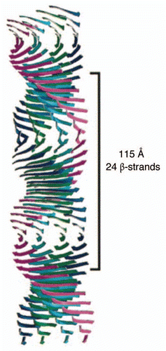
Figure 2 (A) Functional amyloid occurs in different species in different shapes and sizes. Antibody labeling using the amyloid-specific antibody WO1 is shown in green and nuclear counterstaining (DAPI) in blue. (i) C. glutamicum. FuBA is present around all cells. (ii) G. obscurus. FuBA occurs in large extracellular aggregates. The arrows indicate extracellular material with a high level of amyloid but low cell density. Bars 10 µm. (iii) M. avium. Velvet-like substances strongly bind WO2. (iv) T. spumae. Long (up to 50 µm) fibrils (arrows) are present. Bars 10 µm. (B) Saponification of G. amarae at increasing temperatures reveals gradual liberation of fibril-like substances: TEM micrographs with 1% phosphotungstic acid staining of (a) nonsaponified G. amarae, (b) bacteria saponified for 4 days at 37°C, (c) bacteria saponified for 4 days at 60°C and (d) bacteria saponified for 4 days at 80°C. Bars in (a–c) represent 0.5 µm; the bar in d represents 100 µm. The arrows indicate the positions of (a) a dense extracellular matrix and (b to d) fibrillar material. Reprinted with permission from Jordal PB, et al.Citation24
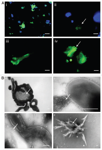
Figure 3 Filamentous Chloroflexi (green) are often covered by epiphytic “Candidatus epiflobacter sp.” (Saprospiraceae, Bacteroidetes, colored yellow) which are specialized in protein degradation.Citation41 Figure provided courtesy of Per Halkjær Nielsen.
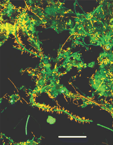
Figure 4 Colony morphotypes of bacterial strains when grown on Congo red agar plates. Bacteria were grown for 48 h at 26°C. It was subsequently possible to purify and identify amyloid from the strain designated UK4.
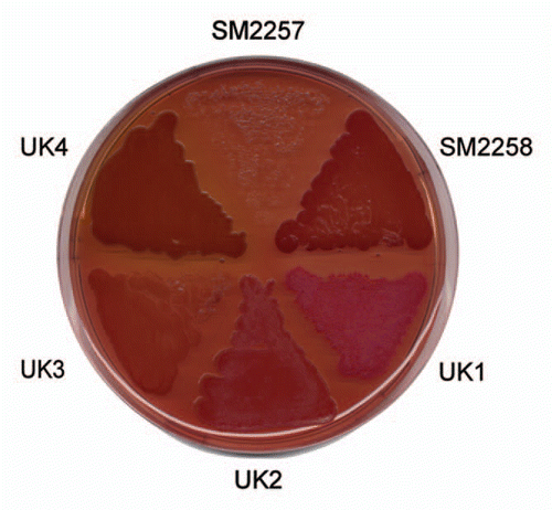
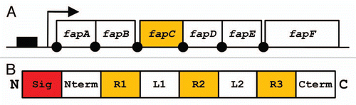
Figure 5 (A) Organization of the fap operon. Promoter region indicated as solid box, protein coding sequences indicated by open boxes. Arrow indicates transcription start site and solid dots are potential ribosomal binding sites. (B) Schematic representation of FapC. The N-terminal signal sequence (Sig) is cleaved off during translocation to the outer membrane, an N-terminal domain (Nterm), three repeats (R1–3) separated by two linker regions (L1–2) and a C-terminal tail (Cterm).