
Abstract
The biochemical essence of prion replication is the molecular multiplication of the disease-associated misfolded isoform of prion protein (PrP), termed PrPSc, in a nucleic acid-free manner. PrPSc is generated by the protein misfolding process facilitated by conformational conversion of the host-encoded cellular PrP to PrPSc. Evidence suggests that an auxiliary factor may play a role in PrPSc propagation. We and others previously discovered that plasminogen interacts with PrP, while its functional role for PrPSc propagation remained undetermined. In our recent in vitro PrP conversion study, we showed that plasminogen substantially stimulates PrPSc propagation in a concentration-dependent manner by accelerating the rate of PrPSc generation, while depletion of plasminogen, destabilization of its structure, and interference with the PrP-plasminogen interaction hinder PrPSc propagation. Further investigation in cell culture models confirmed an increase of PrPSc formation by plasminogen. Although molecular basis of the observed activity for plasminogen remain to be addressed, our results demonstrate that plasminogen is the first cellular protein auxiliary factor proven to stimulate PrPSc propagation.
Figures and Tables
Figure 1 The role of plasminogen in PrPSc propagation. The effect of plasminogen (Plg) was assessed by PMCA using normal brain material supplemented with or without 0.5 µM human Glu-Plg (A–D) or using Plg-deficient (Plg-/-) brain material (F and G). Pre- (−) and post- (+) PMCA samples were treated with proteinase K (PK) and analyzed by western blotting. Seeds for PMCA were diluted either as indicated or 1:900 (G) −8,100 (F). (A) Stimulation of PrPSc propagation by Plg. (B) Plg-supplemented PMCA in the absence of SBH seeds. (C) Plg-supplemented PMCA in the absence of NBH. (D) Comparison of PrPSc levels in Plg-supplemented PMCA samples (during) vs. PMCA samples only incubated with Plg prior to PK digestion (after). (E) Comparison of PrPSc levels of ScN2a cell lysate after incubation with or without Plg prior to PK digestion. (F) PMCA with brain material of Plg-/- mice and genetically unaltered littermate controls (C). (G) Restoration of PMCA using Plg-/- brain material with Plg-supplementation. NBH, normal brain homogenate; SBH, sick brain homogenate; PrPKOBH, brain homogenate of PrPC-deficient mice; CL, cell lysate. Reproduced with permission from The FASEB Journal, Mays and Ryou 2010.Citation35
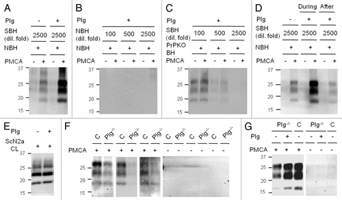
Figure 2 PrPSc propagation increased by plasminogen in prion-infected cells. (A) The levels of PrP in ScN2a cells incubated with 0–0.5 µM human Glu-plasminogen (Plg) for two days. (B) The levels of PrP in ScN2a cells incubated with 0, 0.1 and 1.0 µM Plg or the first three kringle domains of Plg [K(1+2+3)] for six days. (C) The levels of 3F4-tagged PrPC and nascent PrPSc formation in ScN2a cells transiently transfected (Tfx) with plasmids encoding the 3F4-tagged PrP gene (PrP-3F4) and with an empty vector (mock). The transfected cells were treated with 0 or 1 µM K (1+2+3) for three days. (D) The levels of PrP in Elk21+ cells incubated with 0 and 0.5 µM Plg for two days. PrP was detected by anti-PrP antibody D13 (A and B), 3F4 (C) or 6H4 (D) before (−) and after (+) PK treatment. Reproduced by permission of the The FASEB Journal, Mays and Ryou 2010.Citation35
![Figure 2 PrPSc propagation increased by plasminogen in prion-infected cells. (A) The levels of PrP in ScN2a cells incubated with 0–0.5 µM human Glu-plasminogen (Plg) for two days. (B) The levels of PrP in ScN2a cells incubated with 0, 0.1 and 1.0 µM Plg or the first three kringle domains of Plg [K(1+2+3)] for six days. (C) The levels of 3F4-tagged PrPC and nascent PrPSc formation in ScN2a cells transiently transfected (Tfx) with plasmids encoding the 3F4-tagged PrP gene (PrP-3F4) and with an empty vector (mock). The transfected cells were treated with 0 or 1 µM K (1+2+3) for three days. (D) The levels of PrP in Elk21+ cells incubated with 0 and 0.5 µM Plg for two days. PrP was detected by anti-PrP antibody D13 (A and B), 3F4 (C) or 6H4 (D) before (−) and after (+) PK treatment. Reproduced by permission of the The FASEB Journal, Mays and Ryou 2010.Citation35](/cms/asset/f7dff541-457f-4713-b345-58ccb5c01505/kprn_a_10914460_f0002.gif)
Figure 3 Plausible mechanisms for plasminogen to enhance PrPSc propagation. Plasminogen may stimulate PrPSc propagation via conformational alteration of PrPC to PrP* (i), enhancement of PrPSc aggregation (ii), stabilization of pre-exisiting PrPSc aggregates (iii) or scaffolding to gather PrPC and PrPSc together (iv).
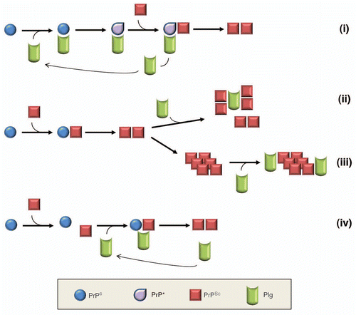
Table 1 Properties of plasminogen as an auxiliary factor for PrPSc propagation