Abstract
The intricate complexity, at the molecular and cellular levels, of the processes leading to the development of amyloid proteinopathies is somehow counterbalanced by their common, universal structural basis. The later has fueled the quest for suitable model systems to study protein amyloidosis under quasi-physiological conditions in vitro and in simpler organisms in vivo. Yeast prions have provided several of such model systems, yielding invaluable insights on amyloid structure, dynamics and transmission. However, yeast prions, unlike mammalian PrP, do not elicit any proteinopathy. We have recently reported that engineering RepA-WH1, a bacterial DNA-toggled protein conformational switch (dWH1 → mWH1) sharing some analogies with nucleic acid-promoted PrPC → PrPSc replication, enables control on protein amyloidogenesis in vitro. Furthermore, RepA-WH1 gives way to a non-infectious, vertically-transmissible (from mother to daughter cells) amyloid proteinopathy in Escherichia coli. RepA-WH1 amyloid aggregates efficiently promote aging in bacteria, which exhibit a drastic lengthening in generation time, a limited number of division cycles and reduced fitness. The RepA-WH1 prionoid opens a direct means to untangle the general pathway(s) for protein amyloidosis in a host with reduced genome and proteome.
Acknowledgments
We apologize to the colleagues whose relevant work we have not quoted due to strict space limitations. Research in our laboratory on RepA-WH1 amyloids is currently financed by Spanish MICINN grants BIO2009-06952 and CSD2009-00088.
Figures and Tables
Figure 1 The bacterial RepA-WH1 prionoid highlighted at the molecular and cellular levels. The stable, soluble dimers of the isolated RepA-WH1 domain (dWH1, 1HKQ.pdb) undergo a conformational transformation upon transient, low affinity (albeit sequence-specific) binding to dsDNA ligand, thus resulting in metastable, aggregation-prone monomers (mWH1, modeled on 1REP.pdb). The roughly invariant (rms deviation for 76 Cα atoms: 1.34 Å) WH fold is depicted in cyan, whereas segments showing a significant structural shift are shown in blue. The amyloidogenic peptide L26VLCAVSLI34 (C-terminal 2/3 of α2),Citation21 is colored in red, with the side-chain of the hyper-amyloidogenic mutant residue V31 (bold) rendered as CPK spheres. A possible pathway for the exposure of the amyloidogenic stretch is sketched (dashed grey arrows 1–3): (1) DNA binding to dWH1 results in a bend in the β-hairpin (β2–β3),Citation17 which carries the sensor Arg residues (R78, R81, R91 and R93; yellow),Citation22 thus disrupting the dimerization interface (second dWH1 subunit in grey). (2) Simultaneously, β2–β3 pushes the N-terminal α1 that swingsCitation16 and partially unfolds. (3) The opening of such α1 latch releases C-terminal α5 which, in the whole mRepA, will then pack with the N-terminus of the WH2 domain to build an antiparallel β-sheet,Citation17 but in the isolated mWH1 is unfolded (dashed coil) to leave the amyloidogenic stretch largely exposed to the solvent and ready to refold and assemble into an amyloid cross-β sheet.Citation21 Models rendered with PyMOL (www.pymol.org).
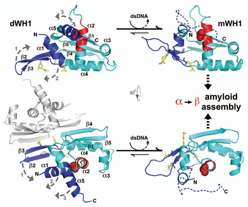
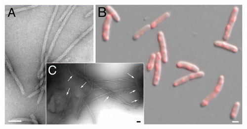
Figure 2 Linking propagation of the bacterial RepA-WH1 prionoid in vitro and in vivo. (A) Electron micrograph of amyloid fibers assembled in vitro by RepA-WH1(A31V) in the presence of effector dsDNA molecules.Citation21,Citation22 Magnification bar: 0.1 µm. (B) E. coli K-12 expressing mCherry-tagged RepA-WH1(A31V) accumulate multiple red fluorescent, aggregated amyloid foci which severely hamper cell proliferation.Citation25 Magnification bar: 1 µm. (C) Molecular transmissibility, the ability of purified bacterial amyloid inclusions/seeds (arrows) to template the transformation and assembly into fibers of soluble RepA-WH1(A31V) molecules,Citation25 links together amyloidosis in vitro and in vivo. Magnification bar: 0.1 µm.