
Abstract
The aerial organs of plants are covered with a cuticle, a continuous layer overlaying the outermost cell walls of the epidermis. The cuticle is composed of two major classes of the lipid biopolymers: cutin and waxes, collectively termed cuticular lipids. Biosynthesis and transport of cuticular lipids occur predominantly in the epidermis cells. In the transport pathway, cuticular lipids are exported from their site of biosynthesis in the ER/plastid to the extracellular space through the plasma membrane and cell wall. Growing evidence suggests that ATP-binding cassette (ABC) transporters are implicated in transport of cuticular lipids across the plasma membrane of epidermal cells. The Arabidopsis ABC-type transporter protein CER5 (WBC12) was reported to act as a wax monomer transporter. In recent works, our group and others showed that a CER5-related protein, DESPERADO (DSO/WBC11), is required for cutin and wax monomers transport through the plasma membrane of Arabidopsis epidermis cells. Unlike the cer5 mutant, DSO loss-of-function had a profound effect on plant growth and development, particularly dwarfism, postgenital organ fusions, and altered epidermal cell differentiation. The partially overlapping function of CER5 and DSO and the fact that these proteins are half-size ABC transporters suggest that they might form a hetero-dimeric complex while transporting wax components. An intriguing observation was the polar localization of DSO in the distal part of epidermis cells. This polar expression might be explained by DSO localization within lipid rafts, specific plasma membrane microdomains which are associated with polar protein expression. In this review we suggest possible mechanisms for cuticular lipids transport and a link between DSO function and polar expression. Furthermore, we also discuss the subsequent transport of cuticular constituents through the hydrophobic cell wall and the possible involvement of lipid transfer proteins in this process.
Addendum to: Panikashvili D, Savaldi-Goldstein S, Mandel T, Yifhar T, Franke RB, Höfer R, Schreiber L, Chory J, Aharoni A. The Arabidopsis DESPERADO/AtWBC11 transporter is required for cutin and wax secretion. Plant Physiol 2007; 145:1345-60.
Figures and Tables
Figure 1 Transport mechanisms involved in cuticular lipids transport in intracellular and extracellular spaces. TMD and NBF-transmembrane domain and nucleotide binding fold of ABC transporter. FABP-fatty acid binding protein. One possibility is that cuticular lipids upon biosynthesis in the ER or plastid are relocated to the PM localized ABC transporter with the aid of FABP. Second possibility is the transport within the oleosome bodies with subsequent incorporation into the PM lipid rafts. DSO and CER5 might interact in the transport of wax monomers, however, additional ABC transporter might form heterodimer with DSO in the delivery of cutin monomers from intracellular space to the outer surface. From the PM cuticular lipids pass through the cell wall to the polymerization site or they are taken up by the lipid transfer proteins (LTPs).
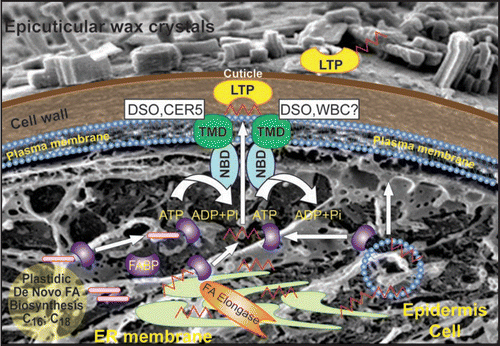
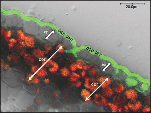
Figure 2 Localization of DSO-GFP protein fusion. Confocal microscopy of stem cross-sections of plants harboring the promoter pDSO::GFP-DSO construct. Ep—indicates epidermal cells and cor—cortex cells.
Addendum to: