
Abstract
Glucose functions in plants both as a metabolic resource as well as a hormone that regulates expression of many genes. Arabidopsis hexokinase1 (HXK1) is the best understood plant glucose sensor/transducer, yet we are only now appreciating the cellular complexity of its signaling functions. We have recently shown that one of the earliest detectable responses to plant glucose treatments are extensive alterations of cellular F-actin. Interestingly, AtHXK1 is predominantly located on mitochondria, yet also can interact with actin. A normal functioning actin cytoskeleton is required for HXK1 to act as an effector in glucose signaling assays. We have suggested that HXK1 might alter F-actin dynamics and thereby influence the formation and/or stabilization of cytoskeleton-bound polysomes. In this Addendum, we have extended our initial observations on the subcellular targeting of HXK1 and its interaction with F-actin. We then further consider the cellular context in which HXK1 might regulate gene expression.
Acknowledgements
Technical contribution no. 5388 of the Clemson University Experiment Station.
Figures and Tables
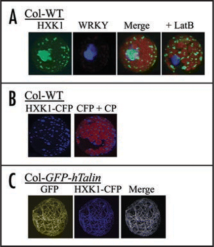
Figure 1 Fluorescence images after transfection of different cDNAs into mesophyll protoplasts. Images were collected using a Zeiss LSM 510 confocal laser scanning microscope and band pass filters for GFP and CFP, and a long-pass filter for chlorophyll. (A) Influence of transient overexpression and actin disruption on sub-cellular targeting of AtHXK1-GFP in Arabidopsis protoplasts. Fluorescence of HXK1-GFP or co-transfected WRKY-CFP was visualized after 12 h expression in protoplasts, without or with 2 µM Latrunculin B. The left 3 images are of the same protoplast not treated with LatB, while the image on the far right is of a different protoplast that was treated with LatB. Note that the light-blue image is due to aggregated GFP and does not specifically overlap with the image from CFP fluroscence. (B) Subcellular targeting of transfected AtHXK1-CFP in leaf protoplasts from Col WT. Shown are fluorescence images of transfected HXK1-CFP in Col WT protoplasts, without (left) or with (right) chlorophyll autofluorescence. Note the punctate distribution of blue fluorescence. This pattern is typical for the mitochondrial targeted HXK1.Citation9 CP = chloroplasts. (C) Subcellular targeting of transfected AtHXK1-CFP in leaf protoplasts from Col GFP-hTalin. Shown from left to right are images of wavelength specific fluorescence for GFP-hTalin bound to F-actin, for transfected HXK1-CFP, and for the merged image. Note that the transfected HXK1-CFP now localizes predominantly to F-actin. In the absence of transfected HXK1-CFP, no corresponding blue wavelength fluorescence was observed in the leaf protoplasts.
Addendum to: