
Abstract
Plants respond to almost any kind of external stimulus with transients in their cytoplasmic free calcium concentration ([Ca2+]c). A huge variety of kinetics recorded by optical techniques has been reported in the past. This variety has been credited the specificity needed to explain how information about incoming stimuli is evaluated by the organism and turned into the right physiological responses which provide advantages for survival and reproduction. A physiological response often takes place away from the site of stimulation. This requires cell-to-cell communication. Hence, responding cells are not necessarily directly stimulated but rather receive an indirect stimulus via cell-to-cell communication. It appears unlikely that the '[Ca2+]c signature' in the primarily stimulated cell is conveyed over long distances via cell-to-cell communication from the 'receptor cells' to the 'effector cells'. Here, a novel aspect is highlighted to explain the variety of [Ca2+] kinetics seen by integrating methods of [Ca2+]c recording. Plants can generally be seen as cellular automata with specific morphology and capable for cell-to-cell communication. Just a few rules are needed to demonstrate how waves of [Ca2+]c-increases percolate through the organism and thereby deliver a broad variety of 'signatures'. Modeling intercellular signaling may be a possible way to find explanations for different kinds of signal transmission, signal amplification, wave formation, oscillations and stimulus-response coupling. The basic examples presented here show that care has to be taken when interpreting cellular '[Ca2+]c signatures' recorded by optical techniques which integrate over a big number of cells or even whole plants.
Acknowledgements
I thank Hartmut Kaiser (Botanical Institute, University Kiel) for critically reading the manuscript. The author received financial support from the DFG and the federal state of Schleswig-Holstein.
Figures and Tables
Figure 1 A single cell response after stimulation. The cell was stimulated at t = 50. A timescale of 1: = 0.1 sec gives a realistic estimate of the real kinetic in a living plant. The parameters according to EquationEq. 1 for this [Ca2+]c-spike are: .
![Figure 1 A single cell response after stimulation. The cell was stimulated at t = 50. A timescale of 1: = 0.1 sec gives a realistic estimate of the real kinetic in a living plant. The parameters according to EquationEq. 1[Ca2+]c(t)=[Ca2+]c0+a1⋅(1−exp(−tτE))+a2⋅(1−exp(−tτR)) for this [Ca2+]c-spike are: Table 1.](/cms/asset/ed9c48af-ec68-4876-a392-7dcf7bf9618c/kpsb_a_10910717_f0001.gif)
Figure 2 The signal percolation in a two cell system. (A) The response in the second cell (blue trace) occurs when the [Ca2+]c in the stimulated cell exceeds half maximum (a level of [Ca2+]E = 400 nM). This results in a delay time of Δt = 5. (B) The [Ca2+]c traces of both cells overlap in time. It is obvious that the sum (=overall signal of the two-cell system) does not differ significantly from the single cell response.
![Figure 2 The signal percolation in a two cell system. (A) The response in the second cell (blue trace) occurs when the [Ca2+]c in the stimulated cell exceeds half maximum (a level of [Ca2+]E = 400 nM). This results in a delay time of Δt = 5. (B) The [Ca2+]c traces of both cells overlap in time. It is obvious that the sum (=overall signal of the two-cell system) does not differ significantly from the single cell response.](/cms/asset/ab749350-416d-4098-9c00-7591ab8ec6f1/kpsb_a_10910717_f0002.gif)
Figure 3 A presentation of the four cell system. (A) In a four cell system the configuration can be either a running of the signal from one end through the whole system (left side in A) or from the middle to the borders (right hand side in A). (B) The single responses from the individual cells. (C) The sum of all (bold line) gives the observed signal. It is slightly different from that of a single cell given as gray trace for comparison.
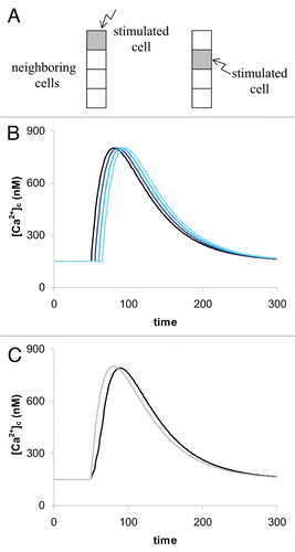
Figure 4 The 16-cells system. (A) The compact system with stimulation in the middle. (B) The elongated system (domino configuration) with stimulation at one end. (C) [Ca2+]c-traces of all 16 cells in the domino configuration. (D) The over-all responses of the whole system in the ‘domino configuration’ (green) and in the compact configuration (pink). This trace is almost the same as in the 4-cell system as shown in . For comparison the response of a single cell is shown (gray line). This is identical with a whole systems response when all cells are simultaneously stimulated.
![Figure 4 The 16-cells system. (A) The compact system with stimulation in the middle. (B) The elongated system (domino configuration) with stimulation at one end. (C) [Ca2+]c-traces of all 16 cells in the domino configuration. (D) The over-all responses of the whole system in the ‘domino configuration’ (green) and in the compact configuration (pink). This trace is almost the same as in the 4-cell system as shown in Figure 3C. For comparison the response of a single cell is shown (gray line). This is identical with a whole systems response when all cells are simultaneously stimulated.](/cms/asset/bb5c6e72-434c-4a76-a093-6c8e12e44212/kpsb_a_10910717_f0004.gif)
Figure 5 The 225 cells system. (A) The compact 225 cell configuration (15 × 15 cells). (B) The responses of a 225-cell system in the ‘domino’ configuration (blue), in the compact configuration (green), in the ‘avalanche’-configuration (red). The gray trace represents the response when all cells are responding simultaneously (dotted line) and is identical with the response of a single cell. [Ca2+]E was here set to 310 nM which results in a delay time of Δt = 3. All other model parameters are the same as in . The inset is a close-up and represents the modelled data between t = 40 and 240.
![Figure 5 The 225 cells system. (A) The compact 225 cell configuration (15 × 15 cells). (B) The responses of a 225-cell system in the ‘domino’ configuration (blue), in the compact configuration (green), in the ‘avalanche’-configuration (red). The gray trace represents the response when all cells are responding simultaneously (dotted line) and is identical with the response of a single cell. [Ca2+]E was here set to 310 nM which results in a delay time of Δt = 3. All other model parameters are the same as in Figure 1. The inset is a close-up and represents the modelled data between t = 40 and 240.](/cms/asset/696a1040-d9ed-42be-8b10-4480f469f85c/kpsb_a_10910717_f0005.gif)
Figure 6 Dependence of the overall [Ca2+]c kinetic on the location of the primary stimulus and on the number of primarily stimulated cells in the ‘young-seedling configuration’. (A) All cells of the left cotyledon are stimulated simultaneously; (B) The surface cells of both cotyledons are stimulated; (C) Two meristematic cells are stimulated; (D) The root tip only is stimulated. (E) Dependent on the site of stimulation and the number of primarily stimulated cells, very different [Ca2+]c kinetics are obtained from the whole system.
![Figure 6 Dependence of the overall [Ca2+]c kinetic on the location of the primary stimulus and on the number of primarily stimulated cells in the ‘young-seedling configuration’. (A) All cells of the left cotyledon are stimulated simultaneously; (B) The surface cells of both cotyledons are stimulated; (C) Two meristematic cells are stimulated; (D) The root tip only is stimulated. (E) Dependent on the site of stimulation and the number of primarily stimulated cells, very different [Ca2+]c kinetics are obtained from the whole system.](/cms/asset/9a23ffe7-e0e0-41e4-914f-bf79c81973e9/kpsb_a_10910717_f0006.gif)
Table 1 Model parameters used according to EquationEquation 1 for calculating curves in to