
Abstract
Cytoplasmic pH has long been considered to act as a secondary messenger of various cellular responses by affecting the ionization state of proteins.1 In plant biology, cytoplasmic pH has traditionally been measured, especially in guard cells and as a response to plant microorganism interactions, with pH-sensitive microelectrodes.2 More recently, the development of fluorescent pH markers, such as BCECF and SNARF-1, has allowed us to monitor cytoplasmic pH without the need for electrophysiological equipment. However, because of vacuolar structures that occupy a large volume of plant cells, simple measurements of fluorescent intensities are insufficient to provide precise cytoplasmic pH values. In this addendum, we describe our improved method to monitor cytoplasmic pH in plant cells stained by SNARF-1 by image processing using a noise-reducing filter after determination of an optimal ROI size. In addition, further developments for automated region extraction are proposed.
Figures and Tables
Figure 1 Typical images of tobacco BY-2 protoplasts stained by SNARF-1. (A–D) and (E–H) show images of a cell adjusted to pH 8.0 (Cell A) or pH 7.0 (Cell B), respectively. (A and E), (B and F) and (C and G) show images obtained from channel 1 (Ch1, 540–590 nm), channel 2 (Ch2, 610–670 nm) and the ratio of channel 2 and 1 (Ch 2/1), respectively. (D and H) show images processed by an averaging filter with a radius of 5 pixels in every channel. The box in (H) shows a square region of 10 pixels used for ROI setting. The bar represents 10 µm.
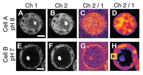
Figure 2 Scatter plots of the image florescent intensities. (A and B) Scatter plots of fluorescent intensities obtained from images in , B, E and F, respectively. (C and D) Scatter plots from images after processing with an averaging filter of 10 pixels in radius.
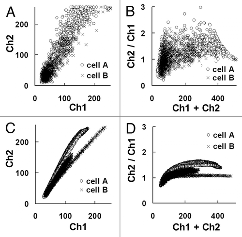
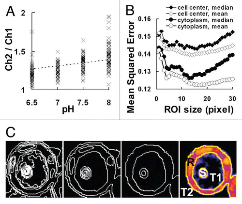
Figure 3 Regression line for pH monitoring and its evaluation by MSE. (A) Scatter plot of the channel ratio in cells adjusted to pH 6.5 to 8.0. The regression line obtained by the least-square method is shown as a dotted-line. (B) Changes in the MSE (mean squared error) of the regression line by the region of interest (ROI) size. The ROI was selected from the cell center or cytoplasm and an averaging or median filter of the indicated ROI size was used in the processing, respectively. (C) Images processed by automatic segmentation of cell B in . From left to right panels, pixels were connected according to their similarity and the image was finally segmented into the four regions of the cytoplasm (R), cell nuclei or nucleus (S), vacuole (T1) and the cell exterior (T2).
Addendum to: