
Abstract
Two sperm cells are required to achieve double fertilization in flowering plants (angiosperms). In contrast to animals and lower plants such as mosses and ferns, sperm cells of flowering plants (angiosperms) are immobile and are transported to the female gametes (egg and central cell) via the pollen tube. The two sperm cells arise from the generative pollen cell either within the pollen grain or after germination inside the pollen tube. While pollen tube growth and sperm behaviour has been intensively investigated in model plant species such as tobacco and lily, little is know about sperm dynamics and behaviour during pollen germination, tube growth and sperm release in grasses. In the March issue of Journal of Experimental Botany, we have reported about the sporophytic and gametophytic control of pollen tube germination, growth and guidance in maize.1 Five progamic phases were distinguished involving various prezygotic crossing barriers before sperm cell delivery inside the female gametophyte takes place. Using live cell imaging and a generative cell-specific promoter driving α-tubulin-YFP expression in the male germline, we report here the formation of the male germline inside the pollen grain and the sperm behaviour during pollen germination and their movement dynamics during tube growth in maize.
Acknowledgements
The German Research Council DFG (Grant DR 334/2-6) is acknowledged for financial support and Dave Jackson from the Maize Cell Genomics project for providing the α-tubulin-YFP marker line.
Figures and Tables
Figure 1 Establishment and properties of the male germline in maize. Germline cells are labeled with α-tubulin-YFP. (A–D) Microspore at late bicellular stage: (A) the vegetative cell still contains a vacuole (V) and the generative cell nucleus (GN) already underwent DNA-syntheses as indicated by its bright signals compared with the vegetative nucleus (VN) after DAPI staining. (B) α-tubulin-YFP expression is restricted to the generative cell (GC). (C) Merged image of (A and B). (D) α-tubulin-YFP forms a cage around the GC nucleus during Pollen Mitosis II . (E) Transition towards tricellular stage: microtubuli bundles have been formed around and between sperm nuclei (SN1/2). Note that first microtubular tail-like extensions are visible (arrow). (F) At early tricellular stage, twin sperm cells (SC1/2) are arranged in parallel. A microtubuli knot (asterisk) becomes visible at half distance between sperm nuclei. (G) Late and mature tricellular pollen stage showing twin sperm cells with long microtubular extensions (arrow), a microtubuli knot (asterisk) connecting both cells and the germination pore (P). (H and I) DAPI staining to show that sperm cells and vegetative nucleus travel as male germ unit (MGU). Note that initially the nucleus of the leading sperm cell (SCVN) seems closely associated with the vegetative nucleus (VN). (I and J) At later stages leading and trailing sperm cells (SCVN and SCUA) are hardly distinguishable as they change positions inside the growing pollen tube. Arrows mark tail-like microtubuli extensions. (K) Stretched sperm cells measure up to 155 µm in length. An asterisk labels the position of a microtubuli knot between both sperm cells. (L and M) Examples of enlarged microtubuli knots. (N) A manipulated pollen tube (PT) releases twin sperm cells that become spherical within seconds. (O) Spherical sperm cells have completely lost their microtubular structure. Dark areas inside sperm cells are sperm nuclei. Scale bars: 20 µm.
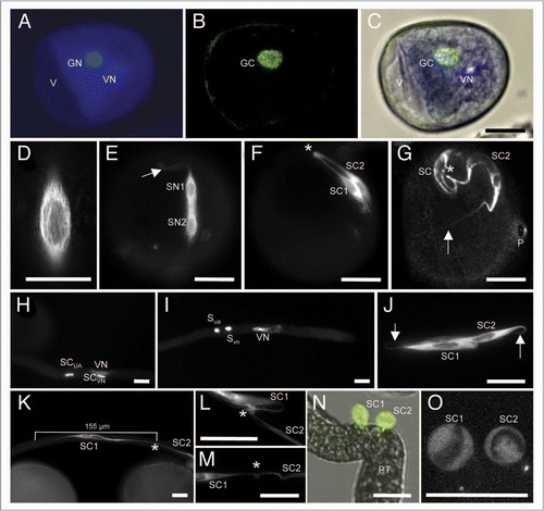
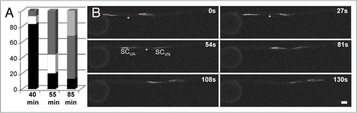
Figure 2 Sperm cell movement into and within the pollen tube of maize. (A) The position of the twin sperm cells was determined at different time points after germination in vitro. Time points of 40 (n = 29), 55 (n = 45) and 85 min (n = 31) are given. Position of sperm cells in percentage is indicated inside the the pollen grain (black bars), while leaving the grain (white bars), in the middle of the pollen tube (dark grey bars) and at the tip of the tube (light grey bar). (B) Time series showing stretched sperm cells (leading and trailing sperm cell SCVN and SCUA, respectively) leaving the pollen grain (left). Movement is shown within a period of 130 seconds (s) as indicated. Please note that movement is very fast after pollen grain exit (top left image) and slows down in the middle of the tube (lower right image). Sperm cells are labelled with α-tubulin-YFP. The tubular knot connecting twin sperm cells is visible in some images (asterisks). Scale bar: 20 µm.
Addendum to: