
Abstract
The respiration rates R (oxygen uptake per min) and body mass M (mg per individual) of sunflower (Helianthus annuus L.) seedlings were measured for populations raised in the dark (scotomorphogenesis) and for plants subsequently grown in white light (photomorphogenesis) to determine the allometric (scaling) relationship for R vs. M. Based on ordinary least squares and reduced major axis regression protocols, cellular respiration rates were found to increase non-linearly as a 'broken-stick' curve of increasing M. During germination, the scaling was ca. 7.5-fold higher than after the emergence of the cotyledons from the seed coat, which can be attributed to the hypoxic conditions of the enclosed embryo. During seedling development, R was found to scale roughly as the 3/7 power of body mass (i.e., R ~ M −3/7), regardless of whether plants were grown in the dark or subsequently in white light. The numerical value of 3/7 statistically significantly differs from that reported across field- or laboratory-grown plants (i.e., R ~ M −1.0). It also differs from the expectations of recent allometric theory (i.e., R ~ M −0.75 - 1.0). This difference is interpreted to be the result of species-specific tissue-compositions that affect the volume fractions of metabolically active and less activ cells. These findings, which are supported by cytological and ultrastructural observations (i.e., scanning- and transmission electron micrographs), draw attention to the need to measure R of developing plants in a tissue- or organ-specific context.
Acknowledgements
We thank Prof. W.R. Briggs (Department of Plant Biology, Carnegie Institution for Science, Stanford, CA) for providing laboratory space and consultation.
This work was supported by the Alexander von Humboldt-Stiftung (AvH, Bonn, Germany, Fellowship Stanford/USA 2009–2010 to UK).
Figures and Tables
Figure 1 Germination (A) and seedling development (B) in sunflower (Helianthus annuus). Achenes were sown in moist vermiculite and raised in darkness (D, days 0 to 2) or grown for 3 days in the dark and subsequently irradiated for 1 to 3 days with continuous white light (WL). The seedlings were kept in 99% relative humidity at 25°C.
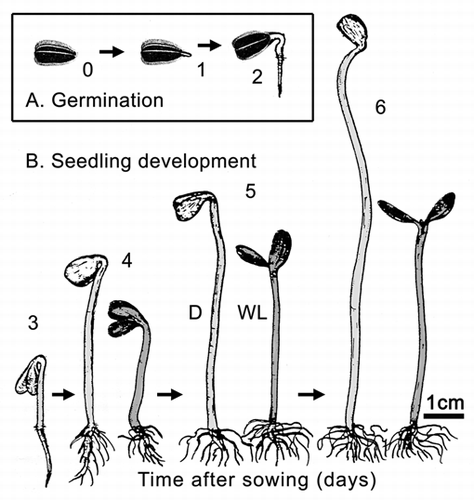
Figure 2 Bivariate plot of log10-transformed data for cellular respiration (R) versus fresh mass (Mf) during germination and seedling development. Sunflower seeds were raised in the dark or, after 3 days of plant growth, irradiated with continuous white light (WL). Lines are reduced major axis (RMA) regression curves (summary statistics provided in the text).
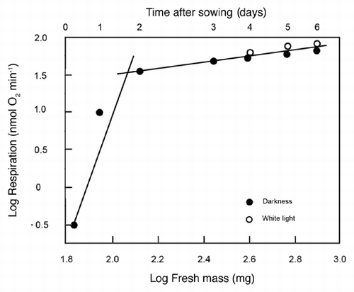
Figure 3 Representative scanning electron micrographs of the cotyledons (A) and epidermal cells (B) of 3-day-old sunflower seedlings that were grown in darkness. The peripheral cells (B) were photographed in the area indicated by the arrow (A). C, cotyledons; E, epidermal cells; S, open stomatum with open pore. Bars = 1 mm (A), 50 µm (B).
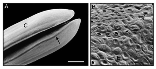
Figure 4 Ultrastructure of sub-epidermal cells in the region between the apical hook and the onset of the cotyledons in 3-day-old etiolated sunflower seedlings. The representative transmission electron micrograph shows the cytoplasmic region of three cells in the periphery of the organ that are separated by a thick wall. A, amyloplast; C, cytoplasm; L, lipid droplet (oleosome); M, mitochondrion; N, nucleus; S, starch; V, vacuole; W, cell wall. Bar, 2 µm.
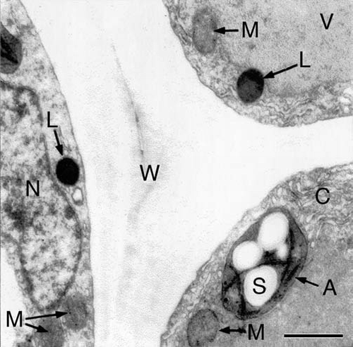
Figure 5 Bivariate plot of log10-transformed data for fresh mass (Mf) versus dry mass (Md) during germination and seedling development. Sunflower seeds were raised in the dark or, after 3 days of plant growth, irradiated with continuous white light (WL). The line is a reduced major axis (RMA) regression curve (summary statistics provided in the text).
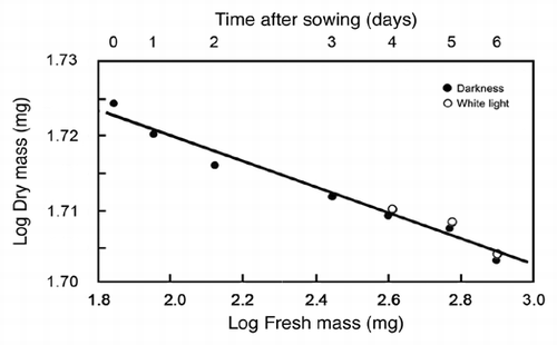
Table 1 List of abbreviations and definitions