
Abstract
Spermatogenesis is a series of cellular processes that leads to the development of motile, elongate sperm cells. Mitotic expansion of spermatogenic stem cells is followed by two meiotic cell divisions that yield haploid round spermatids which then transform from a spherical form into an elongate, highly polarized form. In Drosophila, spermatogenesis takes place within encapsulating cysts that contain spermatogenic cells. Spermatogenic cysts were isolated and grown in culture over the course of 96 hours. Cultures were treated with buthionine sulfoximine (BSO), glutathione (GSH), insulin, and GSH+insulin in order to test the effects of these agents on cyst viability. The addition of glutathione and exogenous insulin to cultured spermatogenic cysts each appeared to have a positive effect on early spermatogenic cyst survival in vitro at some timepoints. The addition of GSH+insulin together had no significant effect on early spermatogenic cyst survival in vitro. Oxidative stress induced by BSO resulted in a significant decrease and/or complete loss of specific early spermatogenic cyst types and the abnormal development of elongating cysts in culture. This culture system offers the opportunity for high-resolution analysis of spermatogenic processes not previously possible.
Acknowledgments
We gratefully acknowledge Allan Blake, Carolyn Bentivegna, Heping Zhou, Monicah Njogu, Crystal Pristell, Zain Alvi and Vikram Basava (SHU) for many helpful discussions, as well as two anonymous reviewers for insightful comments on the manuscript. We gratefully thank Jennifer Goonetilleke and Juan Franco (SHU) for the testes dissections in and B. We thank the SHU Department of Biological Sciences for funding this work.
Figures and Tables
Figure 1 Spermatogenesis in D. pseudoobscura pupal testes. (A) Paired ellipsoid testes (T), seminal vesicles (SV) and accessory glands (AG). The stem cell niche is assumed to be in the apex (A) of the D. pseudoobscura testis based on what is known about the placement of the stem cell niche in D. melanogaster. Sperm coiling occurs in the basal end (B) and mature sperm are stored in the seminal vesicle in adults. Phase contrast image. Bar = 100 µm. (B) Single testis showing enclosed cysts. Arrow indicates elongating cysts (E) which span almost the entire length of the testis as they mature. Brightfield image. Bar = 100 µm. (C) Diagram of spermatogenic cyst maturation in D. pseudoobscura. H, hub cell; CySC, cyst stem cell or cyst progenitor cell; GSC, germline stem cell, G, gonialblast; PS, primary spermatocyte; SS, secondary spermatocyte, RS, round spermatid. The number of spermatogenic cells encapsulated by cyst cells is indicated in parentheses.
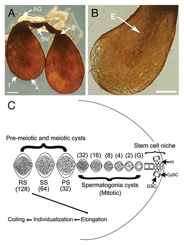
Figure 2 Brightfield images showing spermatogenic cysts after 72 hours in GSH-treated culture. (A) Overall view showing most stages of spermatogenic cyst development. (B) Individualizing cysts with cystic bulges (arrow). (C) Coiling cysts (arrow). Bars = 100 µm. SG, spermatogonia; PS, primary spermatocyte; SS/RS, secondary spermatocyte/round spermatid; E, elongating; IC, individualizing cyst with cystic bulge; CC, coiling cyst; D, degenerating cyst. SS/RS were scored together as it was often difficult to distinguish between them.
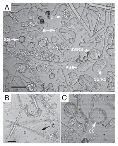
Figure 3 Percentages of each cyst type in pooled data for each treatment type vs. untreated controls. (A) GSH-only, (B) Insulin-only, (C) GSH+insulin, (D) BSO. For each timepoint, the control data bar is on the left and the experimental bar is on the right.
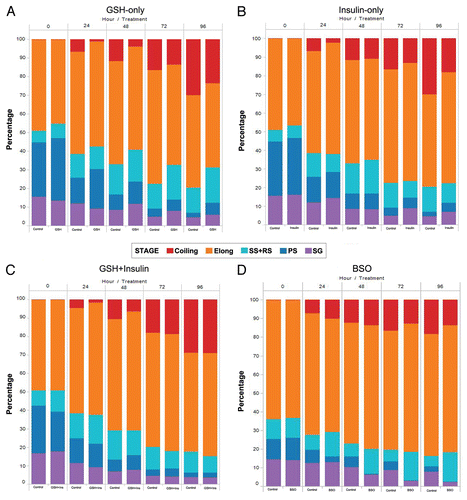
Figure 4 Line graphs showing the change in the raw number of each cyst type for treated and control cultures from the initiation of culture to 96 hours. (A) GSH-only, (B) Insulin-only, (C) GSH-Insulin, (D) GSH/Insulin control, (E) BSO, (F) BSO control.
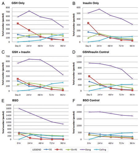
Figure 5 Degeneration counts for primary spermatocytes (PS) in GSH, insulin and GSH+insulin treated cultures. Statistically significant differences are indicated by an asterisk (*). Bars are means ± SEM. *p < 0.05 (Student's t-test). Degeneneration was calculated by determining the total number of lost PS cysts over the course of each 24 hour period then subtracting the increase in secondary and round spermatid cysts. (A) 0 to 24 hours, (B) 24 to 48 hours, (C) 48 to 72 hours, (D) 72 to 96 hours.
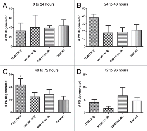
Figure 6 Performance factors for spermatogonia and primary spermatocyte cysts over 96 hours in culture. The ratios are normalized to start at 1 for Day 0 (initiation of culture). Top parts are spermatogonia cysts; bottom parts are primary spermatocyte cysts.
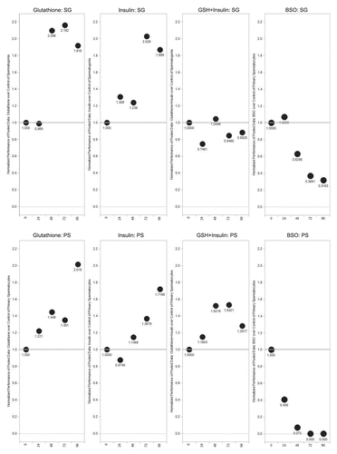
Figure 7 Percentages of spermatogonia and primary spermatocytes in culture vs. untreated controls over 96 hours. Statistically significant differences are indicated by an asterisk (*). Bars are means ± SEM. *p < 0.05 (Student's t-test). (A) GSH-only, (B) Insulin-only. n = 3 for each treatment; n = 5 for untreated controls. (C) GSH+Insulin, (D) BSO. n = 3 for each treatment; n = 5 for untreated controls.
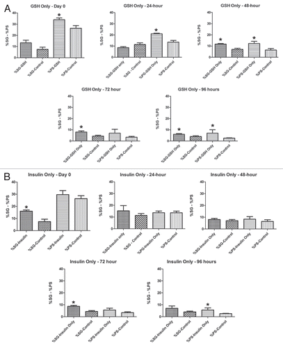
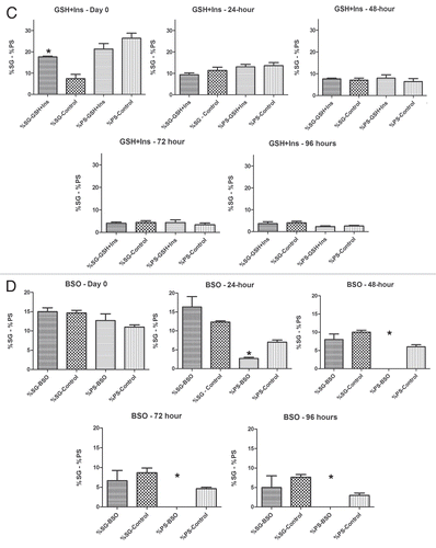
Figure 8 Percentages of early spermatogenic cysts (spermatogonia (SG) + primary spermatocyte (PS)) cysts present over 96 hours in treated and untreated cultures. SG and PS percentages were pooled for GSH, Insulin and GSH+Insulin. (A) Initiation of culture, (B) 24 hours, (C) 48 hours, (D) 72 hours, (E) 96 hours. *p < 0.05 (Student's t-test). Means ± SEM. n = 3 for each treatment; n = 5 for untreated controls.
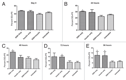
Figure 9 Brightfield images of cultured cysts with fluorescence signal from nuclei (arrows) overlaid. (A) Examples of normal elongating cysts after 24 hours in GSH-treated culture. (B–E) abnormal cysts present after 72 hours in BSO-treated cultures. Several different types of abnormally elongating cysts were observed. In some cases, the nuclei appeared to begin elongation, but became disorganized (large arrows, B and C), while the elongation of the tail portion of the cysts was retarded. Some cysts at the pre-elongation, round spermatid stage also exhibited disorganized nuclei (small arrow, C). (D and E) show cysts with normally elongating nuclei (arrows), but abnormally developed tail regions of the cysts.
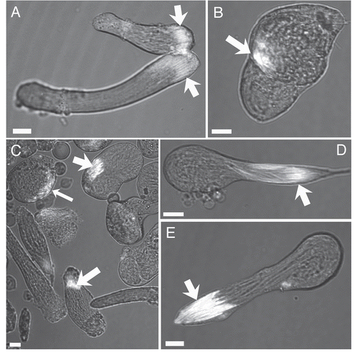
Table 1 Changes in the raw numbers of cysts after 24 hours (0 → 24), 48 (24 → 48), 72 (48 → 72) and 96 (72 → 96) hours