Abstract
Last November a group of principal investigators, postdoctoral fellows and PhD students from around the world got together in the city of Merida in Southeastern Mexico in a State of the Art meeting on the “Molecular structure and function of the apical junctional complex in epithelial and endothelia.” They analyzed diverse tissue barriers including those in the gastrointestinal tract, the blood brain barrier, blood neural and blood retinal barriers. The talks revealed exciting new findings in the field, novel technical approaches and unpublished data and highlighted the importance of studying junctional complexes to better understand several pathogenesis and to develop therapeutic approaches that can be utilized for drug delivery. This meeting report has the purpose of highlighting the results and advances discussed by the speakers at the Merida Meeting.
Introduction
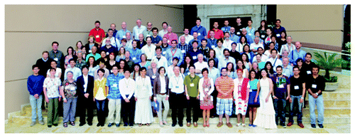
Last November, a couple of days after Hurricane Sandy struck the New York area, the city of Mérida in Southeastern México hosted a meeting where 112 scientists, including postdoctoral fellows and PhD students from around the world, reunited to discuss their latest findings in “Molecular structure and function of the apical junctional complex in epithelial and endothelia” (). Interestingly, while that weekend Mexicans observed the Festival of the Dead, the meeting participants were very much alive. They received more than the sugar skulls or “calaveritas,” as they talked about exciting new findings in the field, novel technical approaches and unpublished data. Here we will highlight some of the results that were discussed in the meeting.
Molecular Structure and Composition of the Apical Junctional Complex
The tetraspan proteins in claudin (Cl) family play a pivotal role in controlling pore properties of tight junctions (TJs) in epithelia and endothelia. Sachiko Tsukita (Kyoto, Japan), a founder of this field who was one of the scientists that discovered this family of proteins described the phenotype of Cl knock out (KO) mice with special emphasis on double Cl−2-/- Cl−15-/- KO mice. These mice have reduced Na+ concentration in the intestinal lumen that results in decreased absorption of glucose, amino acids and fats leading to demise by malnutrition.
Alan Yu’s (Kansas, Missouri, USA) talk highlighted the structure and function of paracellular pores formed by Cl proteins. His studies utilized the first extracellular loop (ECL) of Cl−2 to provide new structural insight on Cl pores that are organized as a hexamer that forms a 6.5 Å diameter cylinder. The model predicts the selectivity of the pore to alkaline metals and is in agreement with experimental values. He went on to demonstrate how cysteine scanning mutagenesis of several amino acids within the ECL1 of Cl−2, followed by a screen to test their accessibility to thiol reactive reagents, is useful to locate and identify the function of individual amino acids within the Cl pore.
Michael Koval (Atlanta, Georgia, USA) spoke of alcoholic lung syndrome in which pulmonary edema is associated with impaired fluid clearance due to increased alveolar leak that correlates with a change in the expression pattern of Cls. Alveolar type I pneumocytes express Cl 4, 5, 7 and 18, while type II pneumocytes additionally express Cl−3. Using an in vivo rat model of alcoholic lung syndrome, Mike observed perturbed expression of Cl proteins with decreased Cl 3, 7 and 18 and increased Cl−5.
Maria Balda (London, UK) spoke of how the guanine exchange factor GEFH1 induces Rho A activation that in turn promotes the nuclear localization of ZONAB transcription factor. This leads to increased cell survival due to the mRNA stabilization of p21, a protein that inhibits the activity of cyclin-CDK1 and two complexes. Since ZONAB is sequestered away from the nucleus by ZO-1, this talk highlighted the relation between TJ adaptor proteins and the regulation of cell cycle progression.
Although James Anderson (Bethesda, Maryland, USA) and his wife and collaborator Christina Van Itallie tried to attend the meeting, Hurricane Sandy disrupted their plans, allowing only his orphan talk to arrive at Merida. Thanks to his commitment to the meeting and the didactic quality of his figures, Asma Nusrat presented his talk where we learned of new methods that his laboratory has employed to examine TJ protein complexes and signaling pathways. These include the use of Phos-tag gel electrophoresis to analyze Cl phosphorylation and use of biotin ligase fused to TJ proteins, followed by biotin administration to biotinylate proteins in close vicinity to TJ bait proteins. The labeled proteins are then captured on a streptavidin affinity resin and identified by mass spectrometry. This systems biology approach will be useful in demonstrating the network of structural and signaling proteins that control TJ function.
Mikio Furuse (Kobe, Japan), who had played a pivotal role in discovery of TJ transmembrane proteins, reported the finding of a new smooth septate junction specific membrane protein named Snakeskin required for the intestinal barrier function. Mikio impressed the audience with impeccable confocal images showing the expression of this protein in Drosophila midgut and Malpighian tubules.
Alan Fanning (Chapel Hill, North Carolina, USA) utilized a deletion approach to demonstrate the role of a TJ cytoplasmic plaque protein, ZO-1. He observed that the SH3 domain and the U5 motif are required to recruit ZO-1 to the apical junctional complex (AJC), PDZ-2 domain is needed to establish a circumferential band of ZO-1, occludin and Cls, and PDZ-1 domain is required for the organization of perijunctional F-actin at the AJC.
Liora Shoshani (Mexico City, Mexico) showed that the β subunit of the Na+-K+-ATPase acts a cell-cell adhesion molecule in the lateral membrane of epithelial cells establishing species specific homophilic trans interactions.
Jerrold Turner’s talk (Chicago, Illinois, USA) addressed the role of occludin phosphorylation (S408) by CK2 in regulating TJ proteins and epithelial barrier function. He observed that the S408A occludin mutant exhibited increased binding of Cl−1 and Cl−2 in comparison to the S408D mutant protein. In addition, CK2 inhibition elevates transepithelial resistance to passive ion flow and inhibits barrier compromise induced by the pro-inflammatory cytokine IL-13 and this effect is mediated by Cl−2.
Organization of Tight Junction Proteins at the Blood Brain Barrier
The unique organization of TJs in the endothelial cells of the blood brain barrier (BBB) is critical in maintaining this structure and for protecting the underlying neuronal tissue. Ingolf Blasig (Berlin, Germany), analyzed a novel application for understanding TJ biology in the BBB by using peptides that recapitulate Cls extracellular segments to promote drug delivery across these barriers. He also addressed mechanisms by which Cl peptides mediate these biological effects.
Anuska Andjelkovic (Ann Arbor, Michigan, USA) talked about cerebral cavernous malformations (CCMs) consisting of vascular sinusoids lined by a thin endothelium with poorly defined TJs. Three proteins referred to as CCM1 to 3 are associated with these malformations that lead to an increased risk of stroke, seizures, epilepsy and headaches. Loss of CCM1 alters localization of the AJC protein β catenin and deletion of CCM2 limits Rho A activation while the task of CCM3 is not well understood. A role in ZO-1 association with the actin binding protein, cortactin, was proposed.
Istvan Krizbai (Szeged, Hungary) discussed mechanisms that regulate carcinoma cell migration across the BBB. He talked about mechanisms by which melanoma cells disrupt TJs of endothelia and migrate through the paracellular pathway and highlighted a role of serine proteases in the process.
Reiner Haseloff (Berlin, Germany) described a strategy for enrichment of junction associated proteins, based on their affinity to Clostridium perfringens enterotoxin (CPE). Immunoblotting and mass spectrometry experiments revealed an enrichment of TJ proteins that specifically associate with a GST-CPE fusion protein.
Signaling at the Apical Junctional Complex
Lorenza Gonzalez-Mariscal (Mexico City, México) described how the intracellular traffic of ZO-2 between the nucleus and the TJ is regulated by nuclear localization and exportation signals and by their modification by phosphorylation and O-GlcNAcylation.
Michael Fromm (Berlin, Germany) spoke of Cls 15, 10a, 10b and 17. He revealed that Cl−15 is highly expressed in the intestine and is located in close proximity to the sodium-glucose symporter SGLT1 and suggested that this is crucial for the recirculation of Na+ necessary for glucose uptake. He explained how the two splice variants of Cl−10 exhibit opposite ionic selectivity as Cl−10b functions as a cation pore and Cl−10a as an anion selective channel. Cl−17 exhibits anion selectivity and is strongly expressed in the proximal renal tubules where it might provide the molecular basis of paracellular chloride transport.
Asma Nusrat (Atlanta, Georgia, USA) highlighted the dynamic nature of TJs and the epithelium itself in the intestine. Her group observed increased Cl−7 protein expression in differentiated intestinal epithelial cells. She discussed approaches to identify transcriptional mechanisms by which Cl−7 protein expression is regulated during epithelial differentiation. Additionally, she talked about Cl−4 that is exclusively expressed in differentiated luminal epithelial cells exhibiting tight barrier properties. Inflammation perturbs TJ proteins thereby compromising barrier function. Some of these effects are mediated by secreted proinflammatory cytokines such as interferon gamma and tumor necrosis factor-a. By using fluorescence recovery after photobleaching, her group is analyzing the influence of proinflammatory cytokines on Cl−4 dynamics in intestinal epithelial cells.
Ruben Gerardo Contreras (Mexico City, México) showed that in MDCK cells, epidermal growth factor (EGF) induces an increase in TER that is accompanied by an enhanced expression of Cl−4 and a decrement of Cl−2. Activation of Erk1/2 regulates the content of Cl−4 while prostaglandin synthesis stimulated by EGF functions as a negative feedback.
Marius Sudol (New York, New York, USA) discussed his novel recent findings on a functional complex between YAP and ZO-2. He showed that the first PDZ domain of ZO-2 forms a stable complex with YAP, binding to its C-terminally located PDZ binding motif. Both endogenous YAP and ZO-2 proteins co-precipitate and co-localize in the nucleus of MCDK, MCF7 and MCF10A cells. YAP overexpression in MDCK cells caused increased proliferation but overexpression of ZO-2 inhibited YAP induced proliferation. It is likely that ZO-2, in addition to being a part of the YAP’s nuclear shuttle, also plays a role in the regulation of transcription of proliferative genes. Marius also reported that his lab is completing the generation of mouse knock-in model in which YAP PDZ binding motif is deleted. The phenotype of the knock-in mouse will shed light on the function of YAP-ZO-2 complex.
Dynamics of the Apical Junctional Complex and Epithelial Polarity
While it is well known that the link between epithelial polarity and intercellular junctions is important in the establishment and maintenance of the epithelial barrier, the underlying mechanisms that mediate these biological effects are still not well understood. A role of the PAR3-aPKC-PAR6 polarity complex in TJ assembly and development of the epithelial apical domain has been reported. Shigio Ohno (Yokohama, Japan) discussed a novel relationship between KIBRA, an upstream regulator of Hippo signaling, PAR3-aPKC-PAR6 and TJ development. Overexpression of the aPKC-binding region of KIBRA disrupts TJs in epithelial cells. High expression of KIBRA in low aPKC-expressing cells decreases aPKC activity, leading to loss of polarity.
Next, Benjamin Margolis (Ann Arbor, Michigan, USA) highlighted the dynamics of an evolutionary conserved polarity protein, Crumbs and TJ formation. Knockdown of Crumbs 3 leads to loss of TJs and expansion of the apical surface. Using flourescence recovery after photobleaching he observed a mobile pool of Crumbs3 in the apical membrane. Additionally, Benjamin discussed the relationship of the transcription factor Snail, Crumbs 3 and epithelial mesenchymal transition. These two talks addressed the important relationship between apical membrane development and TJ formation.
The AJC is structurally and functionally linked to the apical cytoskeleton. Andrei Ivanov (Richmond, Virginia, USA) spoke about the role of actomyosin cytoskeleton in controlling this junctional complex. He discussed the non-redundant roles of cytoplasmic β- and γ-actin isoforms in regulation of epithelial apical junctions and the involvement of β cytoplasmic (β-CYA) and γ-cytoplasmic (γ-CYA) actin isoforms in the biogenesis of adherens junctions (AJ) and TJs in the intestinal epithelium. These studies have addressed unique roles of β-CYA and γ-CYA in regulating the steady-state integrity of the AJC and epithelial barrier function. Additionally, the role of non-muscle myosin II (NM IIA) motor in apical junction biogenesis in vitro and barrier function in vivo using knockout mice was discussed. Junction and actin cytoskeletal dynamics are regulated by the Rho family of GTPases.
Sandra Citi (Geneva, Switzerland) talked about the contribution of TJ cytoplasmic plaque proteins, cingulin and paracingulin to Rho protein regulation and expression of TJ transmembrane Cl family of proteins. Interestingly, epithelia from cingulin knockout mice have increased Cl−2 expression that has been linked to Rho A activity. Additionally, she observed that cingulin and paracingulin control of Cl−2 expression was mediated by transcription factor GATA-4.
Karl Matter (London, UK) further highlighted the contribution of SH3BP1, a GTPase-activating protein for the Rho GTPase family members Cdc42 and Rac, as a regulator of junction assembly and epithelial morphogenesis.
In addition to the barrier function, TJ proteins play an important role in controlling epithelial homeostasis that encompasses cell proliferation, differentiation, migration and regulated shedding. Cl−7 localizes in the lateral membranes of epithelial cells. In a recent study, Yan-Hua Chen (Greenville, North Carolina, USA) observed intestinal pathology, including mucosal ulcerations and epithelial cell sloughing in Cl−7 KO mice. She discussed the relationship of Cl−7 integrin β1 in controlling epithelial cell matrix adhesions. Cl−7 KD cells exhibited reduced β1 integrin protein. Furthermore, Cl−7 co-localized and co-immunoprecipitated with integrin β1. She discussed a novel role of Cl−7 in maintaining epithelial cell-matrix interactions by interacting with b 1 integrin.
Inflammation and the Epithelial/Endothelial Barrier
Leukocytes, and their secreted products, e.g., cytokines and chemokines, influence intercellular junction proteins in endothelial and epithelial cells and thereby regulate their barrier properties. Neutrophil migration across the intestinal epithelium results in increased permeability, tissue damage and disease symptoms. Previous reports indicate that junctional adhesion molecule-like protein (JAML) expressed on neutrophils binds to an epithelial TJ coxsackie-adenovirus receptor (CAR) as one of several steps during neutrophil transepithelial migration. Charles Parkos (Atlanta, Georgia, USA) discussed a novel mechanism by which transmigrating myelomonocytic cells actively shed TJ-binding ligands that alter epithelial barrier function and wound healing, thus contributing to disease pathophysiology. Using new functionally JAML inhibitory antibodies, Parkos group observed that the deleterious effects of JAML released by migrating neutrophils on intestinal epithelial wound repair were inhibited with anti-JAML mAb that specifically blocks JAML-CAR binding.
Leukocyte transmigration across the endothelium occurs predominantly via the paracellular route, though ~11% occurs through the cells (transcellular route). During paracellular migration, junctional proteins such as JAMs and VE cadherin participate in regulating this process. VE-cadherin is of dominant importance for the stability of endothelial junctions. Using a novel in vivo mouse model, Dietmar Vestweber’s (Munster, Germany) talk focused on understanding the mechanisms by which VE cadherin regulates endothelial permeability and leukocyte extravasation. His group used a knock-in mouse expressing VE-cadherin-α-catenin fusion protein to study the dynamics of leukocyte migration and the associated signaling pathways in endothelial cells. Reduced leukocyte extravasation in these mice supports a prominent role of physiological paracellular diapedesis. A role of the vascular endothelial protein tyrosine phosphatase VE-PTP and VE-cadherin, phosphorylation of VE-cadherin and associated signaling proteins in controlling leukocyte movement across the paracellular space was discussed.
In the gastrointestinal epithelium, the transcriptional regulator hypoxia-inducible factor (HIF) acts as an endogenous molecular cue to promote resolution of inflammation in mouse models of disease. Protective influences of HIF are attributable, at least in part, to orchestrated regulation of a barrier protection with the intestinal mucosa. Sean Colgan (Denver, Colorado, USA) observed that isoform-specific HIF-1 and HIF-2 target genes, influence barrier function in fundamental ways. As such, a potential therapeutic paradigm is pharmacological activation of HIF via inhibition of the prolyl hydroxylase enzymes, to harness hypoxia-mediated resolution in intestinal mucosal inflammatory diseases.
Pathogen Induced Signaling Events in the Blood-Brain and Blood-Retinal Barriers
The perineurium in peripheral neurons forms the blood–nerve barrier and protects the nerve. The perineurial barrier is formed by TJ proteins, including Cl−1, Cl−5 and occludin. Although the barrier serves as protection, it also hampers drug delivery of analgesic drugs to the peripheral nerve. Heike L. Rittner (Wuerburg. Germany) discussed novel mechanisms that involve modulation of Cl−1 by perineural administration of Cl−1 peptides to decrease barrier function and promote delivery of analgesic agents to the nerve.
Among the tissue barriers discussed, David A. Antonetti (Ann Arbor, Michigan, USA) talked about the blood-retinal barrier. Endothelial cells that line the vasculature of the retina analogous to that in the blood brain barrier require signals from the neuronal tissue to promote development of TJ protein complexes. His laboratory has utilized mass spectrometry to identify occludin phosphorylation sites in retinal vascular endothelial cells. This analysis led to the identification of two sites that provide distinct roles in cell growth and barrier properties. He discussed a Ser490 occludin phosphorylation site that is responsive to vascular endothelial growth factor (VEGF) and leads to subsequent occludin ubiquitination and endocytosis. Importantly, Ser490 to Ala (S490A) mutants of occludin suppress VEGF induced permeability while occludin ubiquitin chimeric proteins lead to increased basal permeability. This Ser490 phosphorylation is also associated with growth of cells and the same S490A mutants prevent VEGF induced angiogenesis as measured by VEGF induced endothelial tube formation, migration and cell proliferation. His data revealed that occludin phosphorylation contributes to VEGF induced permeability and angiogenesis.
Bacterial proteins influence host junction proteins and this mechanism is crucial in the pathogenesis of disease. Fernando Navarro-Garcia (Mexico City, Mexico) talked about a pathogenic mechanisms by which enteropathogenic E. coli protein, EspF promotes redistribution of TJ proteins during pedestal maturation in epithelial cells. This would then facilitate the invasion of bacteria into the host.
Concluding Remarks
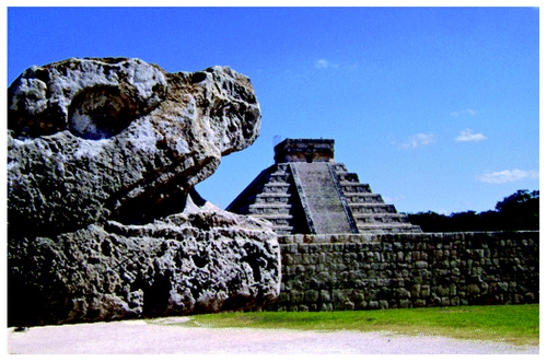
The conference provided a unique forum for scientists studying proteins of the AJC in diverse tissue barrier encompassing epithelia and endothelial in the blood brain barrier, blood neural and blood retinal barriers (). While similarities exist in these systems, the talks highlighted the unique nature of junctional complexes in these diverse sites that cannot only be used to study the biology and disease pathogenesis, but also to develop therapeutic approaches that can be utilized for drug delivery.
| Abbreviations: | ||
| AJ | = | adherens junctions |
| AJC | = | apical junctional complex |
| BBB | = | blood brain barrier |
| CAR | = | coxsackie-adenovirus receptor |
| Cl | = | claudin |
| CPE | = | Clostridium perfringens enterotoxin |
| ECL | = | extracellular loop |
| EGF | = | epidermal growth factor |
| HIF | = | hypoxia inducible factor |
| JAML | = | junctional adhesion molecule-like protein |
| KO | = | knock out |
| KD | = | knock down |
| NM IIA | = | non-muscle myosin II |
| TJ | = | tight junctions |
| VEGF | = | vascular endothelial growth factor |
| β-CYA | = | β cytoplasmic actin |
| γ-CYA | = | γ-cytoplasmic actin |
Acknowledgments
We would like to thank Ingolf Blasig and Reiner Haseloff for their help in the organization of the meeting and insightful ideas. We are also grateful to Liora Shoshani and Gerardo Contreras of the local organizing committee and to the secretary Alicia Teudosio and the students Vicky García and Jorge Lobato for their hard work that made possible this exciting meeting.
Disclosure of Potential Conflicts of Interest
No potential conflicts of interest were disclosed.

