

Abstract
The megaspore genus Biharisporites has a very wide stratigraphic range, being recorded from the Devonian to the Cretaceous. However, in situ these megaspores are known only from the Middle–Upper Devonian, from archaeopteridalean sporangia. Post-Devonian producers of Biharisporites are so far unknown; the parent group can only be hypothesized from the spore morphology and ultrastructure, and from the composition of contemporaneous assemblages of macroremains. To contribute to the understanding of the botanical affinity of dispersed megaspores of the genus, we undertook a comparative ultrastructural study of several Biharisporites species from the Middle Devonian of Russia and the Lower Permian of India. Surprisingly, we found exclusively lycopsid variants of the sporoderm ultrastructure not only in the Permian spores, but also in the Devonian. Therefore, some megaspores of Biharisporites were produced by lycopsids even in the Middle Devonian. Megaspores of Biharisporites morphology have been produced by different groups of spore-bearing plants since the Middle Devonian, and the genus Biharisporites is heterogeneous.
Acknowledgements
We thank Dr. Ludmila I. Kononova (MSU) for providing the Devonian material; Dr. Roman Rakitov (PIN) for his assistance with SEM; Dr. Valentina M. Nazarova (MSU) for advice on the Shchigry-16 borehole lithostratigraphy; Dmitry A. Mamontov (MSU) for maceration of Permian megaspores; and Dr. Maria V. Tekleva (PIN), the editor, and two anonymous reviewers for useful comments and suggestions on the manuscript. The TEM study was carried out at the electron microscopy laboratory of MSU, Biological Faculty, and we are thankful to the team of the laboratory for their assistance. We are also grateful to the administration of Eastern Coalfields Limited, India for granting permission to visit the colliery and help during the field excursion.
Disclosure statement
No potential conflict of interest was reported by the authors.
Plate 1. Images of megaspores of Biharisporites Potonié. All photomicrographs taken with a scanning electron microscope. 1–4, 7–10. Megaspores from the Lower Permian of the Rajmahal Basin, India. 5, 6. Megaspores from the Upper Givetian of the Kursk Region, Russia. 1. Biharisporites cf. spinosus, specimen 409-1, proximal face; the ultrastructure is shown in Plate 3 (figures 1–4), Supplementary Plate S3 (figures 1–3). 2. Biharisporites boralii Bajpai, specimen 409-2, proximal-equatorial view; the ultrastructure is shown in Plate 4 (figures 1–4), Supplementary Plate S4 (figures 1–4). 3. Biharisporites sp. 1, specimen 409-3, proximal face; the ultrastructure is shown in Plate 5 (figures 1–6), Supplementary Plate S5 (figures 1–3). 4. Biharisporites sp. 2, specimen 409-4, proximal face; the ultrastructure is shown in Plate 6 (figures 1–3), Supplementary Plate S5 (figures 4, 5). 5. Biharisporites arcticus var. productus Chi & Hills 1976, specimen 410-03, proximal face; the ultrastructure is shown in Plate 2 (figures 1–5), Supplementary Plate S1 (figures 1–5), and Supplementary Plate S2 (figures 1, 2). 6. Biharisporites arcticus var. productus Chi & Hills, specimen 410-04, distal face. 7. Biharisporites cf. spinosus, enlargement of figure 1 showing the surface of the spore. 8. Biharisporites boralii, enlargement of figure 2 showing the surface of the spore. 9. Biharisporites sp. 1, enlargement of figure 3 showing the surface of the spore. 10. Biharisporites sp. 2, enlargement of figure 4 showing the surface of the spore. Scale bar: 1–6 = 100 µm; 7–10 = 50 µm.
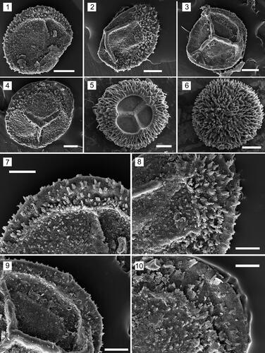
Plate 2. Megaspore ultrastructure of Biharisporites arcticus var. productus Chi & Hills, specimen 410-03 (Upper Givetian of the Kursk Region, Russia), all photomicrographs taken with a transmission electron microscope; the general morphology is shown in Plate 1 (figure 5). 1. Composite image of the section showing bilayered sporoderm, cavity between the layers (c.), gametophyte cavity (g.c.), and the contact area (c.a.); note the intermediate sublayer of the outer layer of the sporoderm (w.o.) that wedges out at the contract area and the apertural region (asterisk). 2. Enlargement of figure 1 showing the apertural region. 3. Fragment of the distal sporoderm, showing sublayers of the outer layer: outer sublayer (o.s.), intermediate sublayer (int.s.), inner sublayer (in.s.), and inner layer (i.l.). The white arrow indicates a sculptural element formed by the outer sublayer of the outer layer. 4. Fragment of the inner layer of the sporoderm showing gaps between the laminae that constitute this layer. 5. Enlargement of figure 2, which clearly shows the laminate structure of the inner layer of the sporoderm, as well as the cavity between the inner and outer layers near the ray of the trilete mark. The black arrow points to the units of the outer layer. Scale bar: 1 = 50 µm; 2, 3 = 10 µm; 4, 5 = 3 µm.
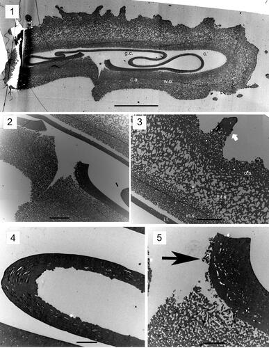
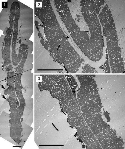
Plate 3. Megaspore ultrastructure of Biharisporites cf. spinosus, specimen 409-1 (Lower Permian of the Rajmahal Basin, India), all photomicrographs taken with a transmission electron microscope; the general morphology is shown in Plate 1 (figure 1). 1. Composite image of the section showing bilayered sporoderm. 2. Enlargement of figure 1 showing the structure of the outer layer. 3. Enlargement of figure 1 showing the gametophyte cavity (g.c.) and the inner layer splitting into a laminated zone (black arrow). 4. Enlargement of figure 3 showing the structure of the inner layer and the laminated zone. Scale bar: 1 = 20 µm; 2, 3 = 5 µm; 4 = 2 µm.
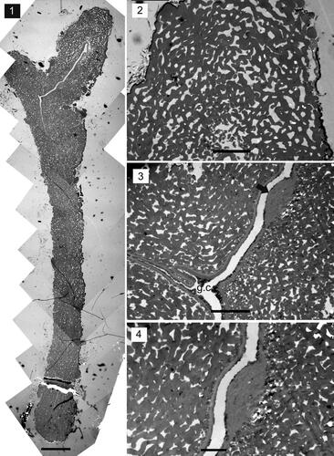
Plate 4. Megaspore ultrastructure of Biharisporites boralii Bajpai, specimen 409-2 (Lower Permian of the Rajmahal Basin, India), all photomicrographs taken with a transmission electron microscope; the general morphology is shown in Plate 1 (figure 2). 1. Composite image of the section showing the arms of the proximal scar (asterisks), inner sublayer (black arrow), and sculptural elements that are composed of the outer layer (black arrowheads). 2. Fragment of the section showing sublayers of the outer layer; the inner sublayer is indicated with a black arrow. 3. Fragment of the sporoderm in the equatorial area; note the inner sublayer (black arrow). 4. The apertural region. Black arrow points to the inner sublayer. Scale bar: 1 = 20 µm; 2–4 = 5 µm.
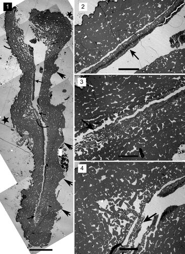
Plate 5. Megaspore ultrastructure of Biharisporites sp. 1, specimen 409-3 (Lower Permian of the Rajmahal Basin, India), all photomicrographs taken with a transmitted electron microscope. 1. Composite image of the section showing the sporoderm, the apertural region (asterisk), and cavities between the inner and outer layers of the distal sporoderm (white arrows) and between the proximal and distal face of sporoderm in the equatorial part of the megaspore (black arrows). 2. Enlargement of figure 1 showing a laminated zone probably cut in its central part (black asterisk). 3. Enlargement of figure 1 showing a laminated zone presumably cut at its periphery (gray asterisk), and the distal (white arrowhead) and the proximal (black arrowhead) parts of the inner layer. 4. The apertural region (asterisk). The black arrow points to a laminated zone presumably cut at its central area, the gray arrow points to a laminated zone presumably cut in its periphery. 5. Enlargement of figure 1 showing thickness of the distal (white arrow) and the proximal (black arrow) parts of the inner layer. 6. Fragment of the section showing the structure of sculptural elements (black arrows). Scale bar: 1, 4 = 20 µm; 2, 5 = 1 µm; 3 = 2 µm; 6 = 10 µm.
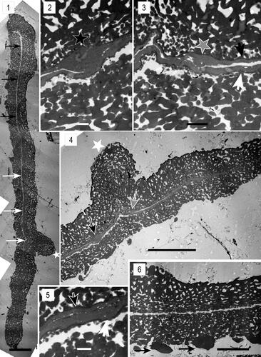
Plate 6. Megaspore ultrastructure of Biharisporites sp. 2, specimen 409-4 (Lower Permian of the Rajmahal Basin, India), all photomicrographs taken with a transmitted electron microscope. 1. Composite image of the section showing the sporoderm and the structure of sculptural elements (black arrows). 2. Fragment of the section. The black arrow points to the inner layer. 3. Fragment of the section showing the structure of the outer layer and the inner layer (black arrow). Scale bar: 20 µm.
Additional information
Funding
Notes on contributors
Alina Kanarkina
ALINA KANARKINA is a master’s student in the Faculty of Geology, Lomonosov Moscow State University, Russia. Her research interests include the ultrastructure of megaspore sporoderms of the Paleozoic higher plants.
Natalia Zavialova
NATALIA ZAVIALOVA is the head of the Laboratory of Paleobotany at the A.A. Borissiak Paleontological Institute, Russian Academy of Science. She graduated from the M.V. Lomonosov Moscow State University, Biological Faculty, where she defended her thesis Morphology and ultrastructure of fossil pollen grains from the upper Permian deposits of Viatka River upstream and the Lower Jurassic deposits of Western Siberia. Her palynological interests include the morphology and ultrastructure of fossil sporoderms from various intervals, such as the Late Paleozoic and Mesozoic. She studies both dispersed and in situ spores and pollen, in the context of their botanical affinities, whole-plant reconstructions, and evolutionary relationships of the parent plant groups. She works for the improvement and wider application of electron microscopy in palynology.
Olga Orlova
OLGA A. ORLOVA is an assistant professor in the Department of Paleontology, Faculty of Geology, at the Lomonosov Moscow State University. She received her PhD for a thesis entitled Visean flora of the Moscow syneclise. Her research interests focus on the morphology and anatomy of the Late Paleozoic fossil plants and megaspores from Central and North Russia.
Arun Joshi
ARUN JOSHI is associated as CSIR-Pool Officer with the Council of Scientific and Industrial Research (CSIR), Government of India. He obtained his PhD from the Birbal Sahni Institute of Palaeosciences, Lucknow, for a thesis titled The Glossopteris flora of Manuguru Area, Godavari Graben: palaeoecological implications, evolutionary perspectives and basinal correlation. His main research interests are Gondwana plant megafossils and megaspores of peninsular and extra-peninsular regions of India.