Abstract
A 57-year-old man developed respiratory symptoms 72 hours after receiving his third dose of the COVID-19 mRNA-1273 vaccine and underwent computed tomography (CT) chest imaging. This showed new diffuse subpleural and peribronchovascular nodular opacities with reverse halo sign. Laboratory investigations demonstrated an eosinophil count of 0.8 x 10E9/L and unremarkable connective tissue disease screen. Bronchoscopy with lavage and transbronchial biopsies demonstrated lymphocytic alveolitis without evidence of infection and histopathology consistent with acute fibrinous and organizing pneumonia (AFOP). He was treated with corticosteroids resulting in resolution of symptoms and CT findings. We present radiographic and histopathologic findings of AFOP following COVID-19 mRNA-1273 vaccination.
Acknowledgments
None.
Disclosure statement
L. Fidler has accepted speaking honoraria from Boehringer Ingelhem, Pfizer and AstraZeneca. The views expressed in the submitted article reflect the authors’ and not an official position of the institution.
Funding
There was no source of financial support for this article.
Figure 1. Computed tomography of the chest post-COVID-19 mRNA-1273 vaccine demonstrating multiple subpleural and peribronchovascular nodular opacities. Most findings show reverse halo sign with peripheral rim of dense consolidation and central ground-glass opacity, more prominent in the lower lung zones.
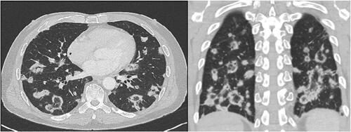
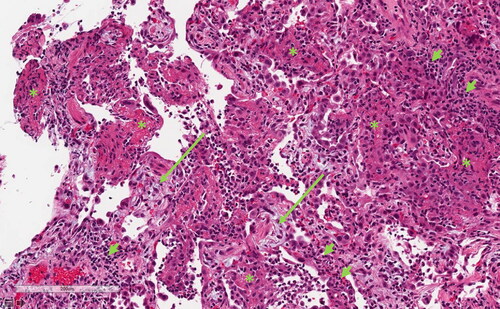
Figure 2. Transbronchial biopsy showing interstitial inflammatory infiltrates (short arrows), with fibrinous exudates filling alveolar spaces (*) and foci of intra-alveolar organizing fibrosis (long arrows). Hematoxylin & eosin stain, scale bar = 200 micrometers.