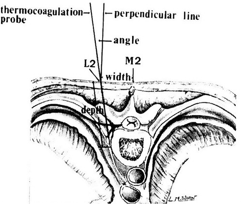
Figure 1 Cross section at the level of second thoracic ganglion shows the entry point (L2), direction and depth of the probe M2 = mid point between the second and third dorsal spinous processes.Citation22
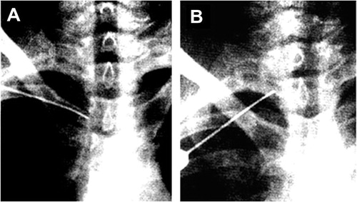
Figure 2 Radiological placement of electrode for RFT of T2 ganglion. (A) The electrode is initially placed adjacent to the T2–T3 interspace. (B) The electrode is placed alongside the T3 vertebral body beneath the head of the second rib.
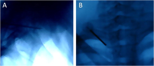
Figure 3 Radiological placement of electrode for RFT of T2 ganglion. (A) Lateral view showing the electrode is placed at the mid vertebral body of T2. (B) P–A view showing the electrode is placed alongside the T3 vertebral body.
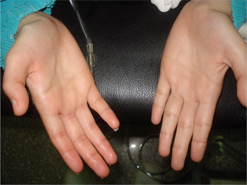
Figure 4 Right palmar flushing and sweating during RFT (stimulation mode) of right T2 ganglion.
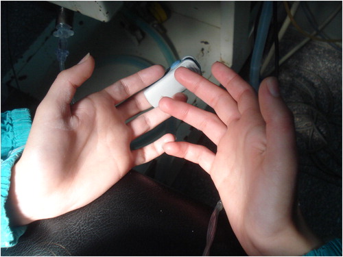
Figure 5 The right hand is dry and flushed after the completion of RFT of right T2 ganglion.
Table 2 Summary data of 10 patients with essential hyperhidrosis who underwent RFT of T2 ganglion.