612
Views
1
CrossRef citations to date
0
Altmetric
Research Articles

Occipitocervical fixation in the management of craniocervical instabilitiesFootnote

&
Pages 185-192 | Received 05 Jun 2011, Accepted 01 Jul 2011, Published online: 17 May 2019

Figures & data

Table 1 Frankel classification of 10 patients with occipitocervical instability.

Table 2 Summary data of 10 patients who underwent occiptocervical fixation.

Table 3 Outcome of patients in relation to the etiology and the technique of fixation.
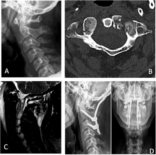
Figure 1 (A) Preoperative plain-Xray cervical spine (lateral view) showing traumatic atlantoaxial sbaxation. (B) Preoperative CT scan (sxial view) showing fracture of both anerior and posterior arch of Atlas. (C) Preoperative MRI T2 weighted image (sagittal view) showing ligamentos injry and swelling of prevertebral tisse at the atlatoxial region. (D) Postoperative plain X-raycervical spine (lateral view) and (axial view) showing ocipitocervical fixation (C0C2C3).
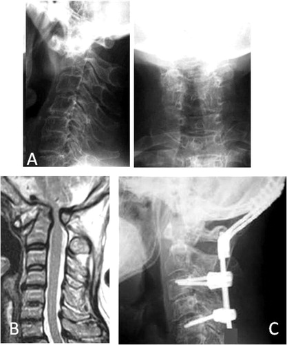
Figure 2 (A) Preoperative plain-Xray cervical spine (lateral view and AP view) of patient with rheumatoid arthritis showing atlantoaxial sublaxation. (B) Preoperative MRI T2 weighted image (sagittal view) showing compression at cervicomedullary junction. (C) Postoperative plain X-ray cervical spine (lateral view) showing occipitocervical fixation (C0C3C5)
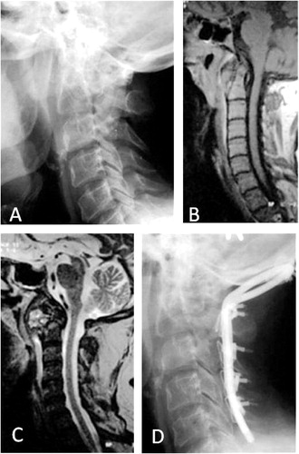
Figure 3 (A) Preoperative plain-Xray cervical spine (lateral view) showing neoplastic destruction of C1 C2 (B) Preoperative MRI T1 weighted image (sagittal view) showing neoplastic mass at C1 C2. (C) Preoperative MRI T2 weighted image (sagittal view) showing neoplastic mass at C1 C2 Postoperative plain X-ray cervical spine (lateral view) showing occipitocervical fixation (C0 C2 C3 C4 C5 fixation) and C1 laminectomy.