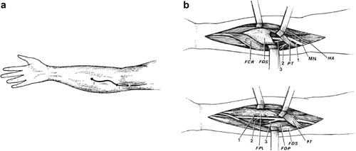
Figure 1 Schematic representation of median nerve neurotomy.Citation3 (a) S-shaped skin incision on the right forearm. (b) Dissection of the median nerve in two stages. First stage of the dissection (upper figure), the PT is retracted upward and laterally and the FCR medially. Branches from the median nerve (MN), before it passes under the fibrous arch of the FDS, are dissected: to the PT(1) and two nerve trunks to the FCR, PL and FDS(2), (3). Second stage of the dissection (lower figure), the fibrous arch of the FDS is sectioned to allow a more distal dissection of the median nerve. The FDS is retracted medially and branches from the median nerve are identified: (1) to the FPL; (2) to the FDP; (3) the interosseous nerve and its proper branches to these muscles.
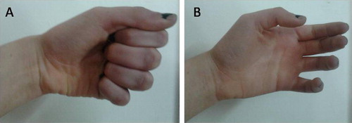
Figure 2 Abnormal spastic hand posture (flexion of wrist and fingers). Before (A) and after (B) surgery.
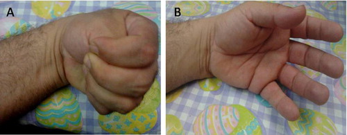
Figure 3 Abnormal spastic hand posture (thumb in hand deformity). Before (A) and after (B) surgery.
Table 3 Summary data and outcome of 10 patients who underwent SPN of median and ulnar nerves.