Figures & data
Figure 1 (A) Sagittal T2 weighted MRI of lumbosacral spine showing decreased normal lumbar lordosis, stenotic bony canal (AP diameter = 8.5 mm at L3/4 and L4/5 levels), diffuse posterior disk bulge at L4/5 together with thickened ligamentum flavum posteriorly (arrow). (B) Axial T2 weighted image at L4/5 level showing diffuse posterior disk bulge slightly inclined to the right side encroaching upon the ipsilateral exiting and traversing nerve roots, exaggerated by bony canal stenosis.
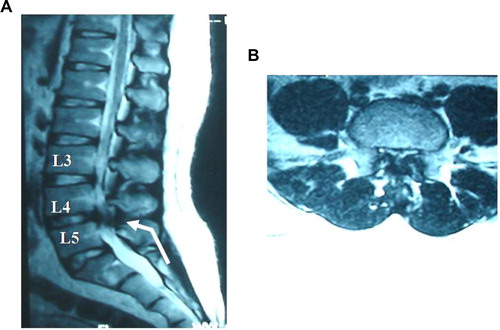
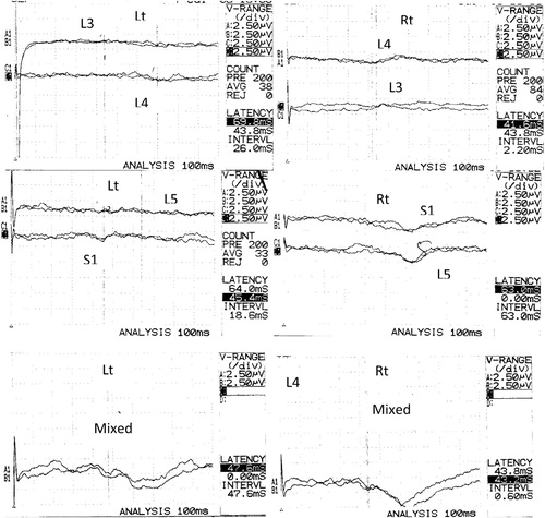
Figure 2 Mixed SEP-PTN and DSEP of L3, L4, L5 and S1 traces of a patient with LSS. Mixed SEP showing normal latency and amplitude on right side stimulation but prolonged latency and low amplitude on left side. At the L3 level, wave forms are absent on the right side but of normal P1 latency and low amplitude on the left side. At the L4 level, normal P1 latency on the right side and prolonged on the left side with low amplitude bilaterally. At the L5 level, the wave forms are absent bilaterally. At the S1 level, also markedly prolonged and attenuated response bilaterally. (R.) right side recording, (L.): left side recording.