986
Views
3
CrossRef citations to date
0
Altmetric
Research Articles

An anatomical study of the blood supply of the soleus muscle in humansFootnoteFootnote

&
Pages 315-321 | Received 31 Jan 2012, Accepted 08 May 2012, Published online: 17 May 2019

Figures & data

Table 1 Distribution of different studied perforators in relation to sex.
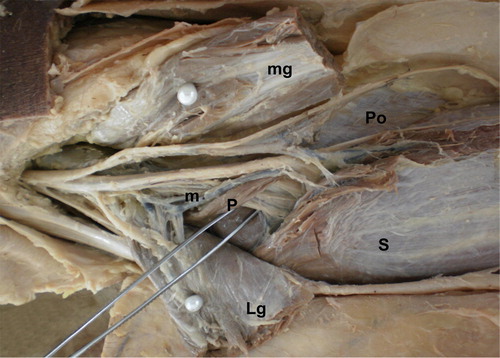
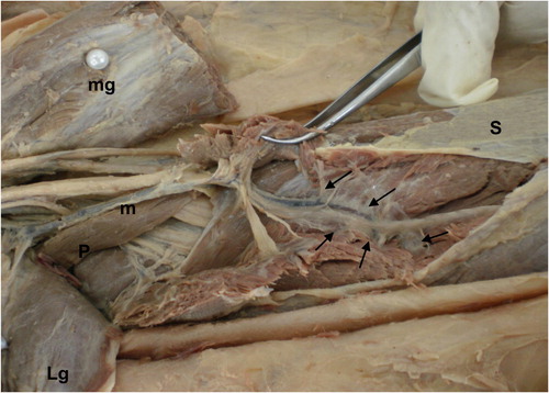
Figure 1 A photograph of the back of the left leg showing the medial (mg) and lateral (Lg) heads of gastrocnemius muscle, they are cut and reflected medially and laterally respectively. The soleus muscle (S) is supplied by a muscular branch (m) that passed through the middle of fits superficial surface and originated from the popliteal artery and supplied the lateral head of gastrocnemius as well. Note the plantaris muscle (P) and popliteus muscle (Po).
Figure 2 A close-up photograph of the previous specimen after further dissection, the muscular branch (m) arising from the popliteal artery in the popliteal fossa passed through the middle of the superficial surface of soleus muscle (S) and then deep into the muscle giving branches to the muscle fibers on the medial and lateral sides (pointed by arrows). Note plantaris muscle (P).
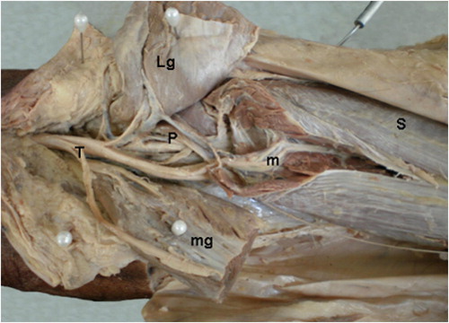
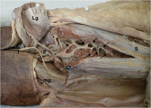
Figure 3 A photograph of the back of the right leg, the lateral (Lg) and medial (mg) heads of gastrocnemius muscles are cut to the show the soleus muscle (S) which is divided through the middle of its superficial surface to show a large muscular branch (m) arising from the popliteal artery and supplying the muscle fibers on both sides. Note plantaris muscle (P) and the branches of the tibial nerve (T).
Figure 4 A photograph of the previous specimen after further dissection showing the muscular branch (m) to soleus (S), it passed through the middle of its superficial surface and then passed deep into the muscle supplying branches to its fibers on both sides (pointed by arrows). Note plantaris muscle (P) and tibial nerve (T).
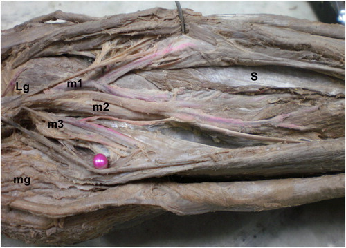
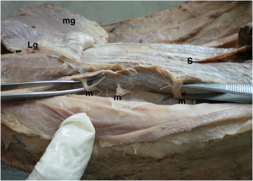
Figure 5 A photograph of the back of the right leg, the two heads of gastrocnemius (Lg and mg) are separated in the midline to show soleus muscle (S) which is divided longitudinally through its middle to show three muscular branches (m1, m2 and m3), arising from the popliteal artery and pass to the lateral, middle and medial parts of the muscle respectively.

Table 2 Comparison between length and diameter of muscular branches of popliteal artery in both male and female.
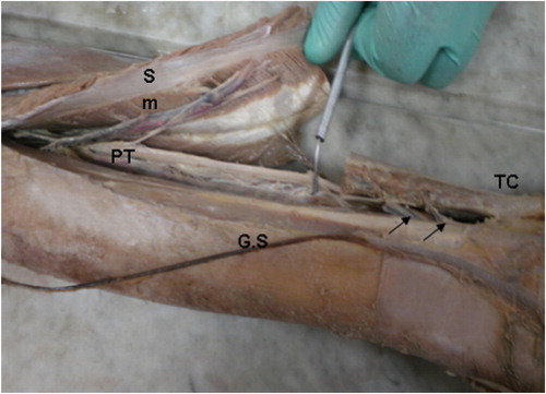
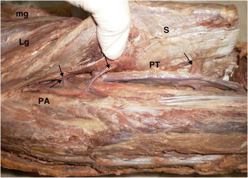
Figure 6 A photograph of the back of the right leg showing the posterior tibial artery (PT) giving a large muscular branch (m) supplying the medial part of soleus muscle (S) through its undersurface, the muscle is cut and its upper part is reflected upwards, its lower part is inserted into tendocalcaneus (TC) and this part of the muscle is supplied by two arteries (pointed by arrows) from the posterior tibial artery. Note the great saphenous vein (G.S).
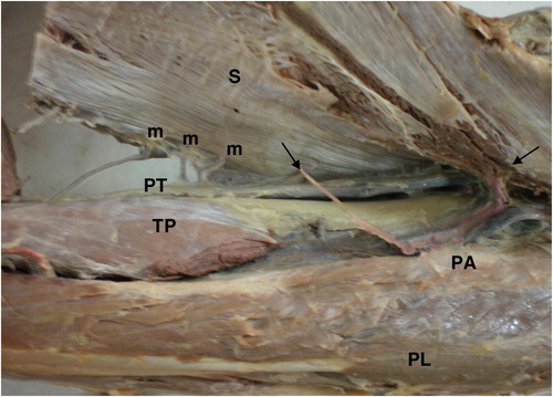
Figure 8 A photograph of the back of the left leg showing the two heads of gastrocnemius muscle (mg and Lg), the soleus muscle (S) which is cut at its lower part and reflected upwards to show the posterior tibial artery (PT) and its peroneal branch (PA) giving muscular branches to soleus muscle (pointed by arrows) that pass through its deep surface.
Figure 9 A photograph of the back of the right leg showing the soleus muscle (S) cut at its lower part and reflected upwards to show the posterior tibial artery (PT) giving muscular branches (m) passing to the medial edge of the muscle. The peroneal artery (PA) gives muscular branches (pointed by arrows) to the deep surface of the muscle. Note peroneus longus muscle (PL) and tibialis posterior muscle (TP).

Table 3 Comparison between length and diameter of muscular branches of posterior tibial artery in both male and female.

Table 4 Comparison between the distance of muscular branches of posterior tibial artery from medial malleolus in male and female in cm.
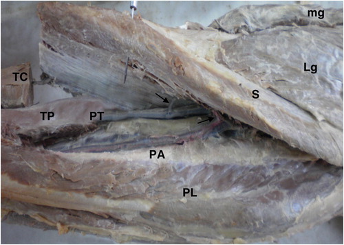
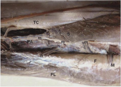
Figure 7 A photograph of the back of the right leg showing the medial (mg) and lateral (Lg) heads of gastrocnemius muscle, the soleus muscle (S) and tendocalcaneus (TC) which is cut and reflected upwards to show the posterior tibial artery (PT) and its peroneal branch (PA) supplying the soleus muscle through its deep surface medially and laterally, respectively, the branches are pointed by arrows. Note the peroneus longus muscle (PL) and tibialis posterior muscle (TP).
Figure 10 A photograph of the back of the right leg showing the peroneal artery (PA) giving muscular branches (m) to soleus muscle (S), nutrient artery (n) to fibula (F) and muscular branch (M) to peroneus longus muscle (PL). Note the tendocalcaneus (TC).
Figure 11 A photograph of the back of the left leg, the two heads of gastrocnemius (Lg and mg) are cut and reflected upwards to show soleus muscle (S). Three muscular branches (m) arise from the peroneal artery and supply the muscle along its lateral edge.

Table 5 Comparison between length and diameter of muscular branches of peroneal artery in both male and female.

Table 6 Comparison between the distance of muscular branches of peroneal artery from lateral malleolus in both male and female in cm.