300
Views
1
CrossRef citations to date
0
Altmetric
Research Articles

Predictive value of brain edema in preoperative computerized tomography scanning on the recurrence of meningiomaFootnoteFootnote

Pages 373-379 | Received 21 Feb 2012, Accepted 02 Jun 2012, Published online: 17 May 2019

Figures & data

Table 1 The age category of patients under study.

Table 2 The gender of patients under study.

Table 3 The number of years since surgery till recurrence in patients under study.

Table 4 The brain edema index in patients.

Table 5 The relationship between the preoperative brain edema index and the average years since surgery to recurrence.

Table 6 The relationship between the preoperative brain edema index and the number of patients with postoperative complications.

Table 7 The most common postoperative complications reported in patients under study.

Table 8 The relationship between the maximum diameter of the tumor and the surrounding brain edema.
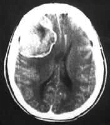
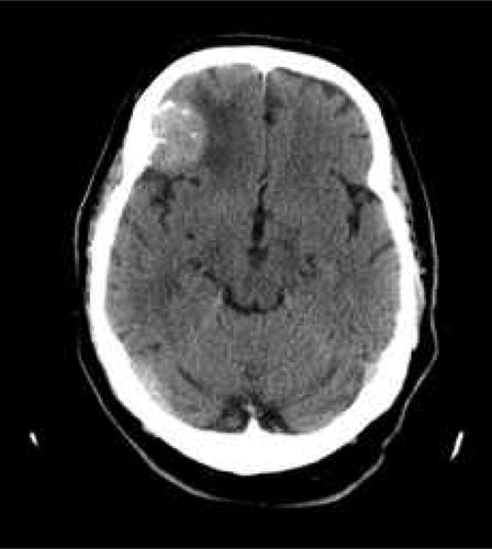
Figure 1 Shows a preoperative axial computerized tomography scan of the brain showing a right frontal convexity meningioma with surrounding brain edema and midline shift.
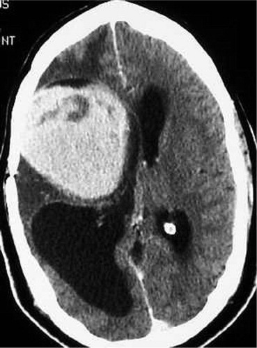
Figure 2 Shows an axial CT scan of the brain, post contrast showing a right frontal convexity meningioma with surrounding brain edema.
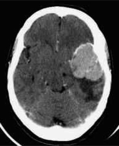
Figure 3 Shows an axial CT scan of the brain, post contrast showing a left sphenoid ridge meningioma with surrounding brain edema.
Figure 4 Shows an axial CT scan of the brain, post contrast that shows a large recurrent right convexity meningioma with surrounding brain edema.
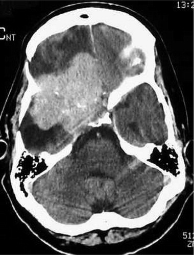
Figure 5 Shows an axial CT scan of the brain, post contrast showing a recurrent right sphenoid ridge meningioma with surrounding brain edema.

Table 9 The relationship between the site of the tumor and the number of cases with brain edema.

Table 10 The correlation between the histopathological subtype of meningioma, preoperative brain edema index, ease of tumor excision and postoperative complications.