Figures & data
Table 1 Different age groups of the study candidates.
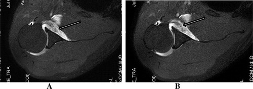
Figure 1 MR arthrogram axial T1w fat sat images of Perthes lesion show non displaced torn segment of the anterior labrum (arrow in A) with still intact glenoid peiosteal link (arrow in B).
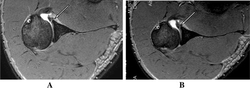
Figure 2 Perthe’s lesion in a 35 year old patient, with minimally displaced inverted torn labral segment(arrow), with still intact periosteum.
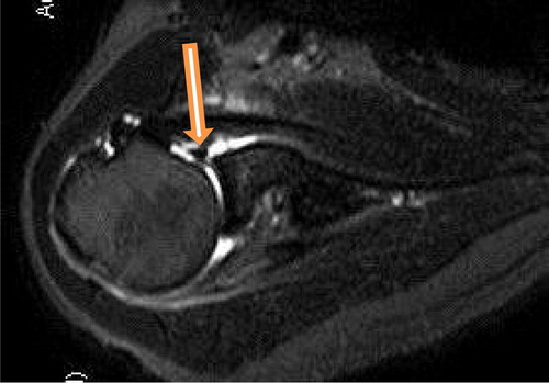
Figure 3 (A, B) Axial T1 fat sat MR arthrogram shows torn and medially displaced anterior glenoid labrum (Arrow in A), with still intact periosteum (Chevron in B), consistent with ALPSA lesion.
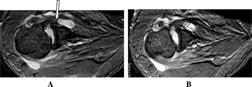
Figure 4 (A, B) Axial MR arthrogram T1 Fat Sat. Images show anterior scapular articular cartilage thin disruption consistent with GLAD injury (arrows).
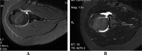
Figure 5 (A, B) Sagittal T1w Fat Sat. MR arthrogram images showing rotator interval tear, seen as extrasynovial leakage of contrast through the defect (arrows) into anterior extra-articular tissues.
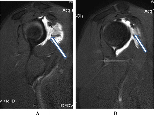
Figure 6 (A) Diagram of type II and III normal variants of labral attachment, as shown in coronal MRI sequence (arrows).Citation20 (B, C) Coronal MR arthrogram T1 fat sat. Images showing SLAP II injury of superior labrum with non-detached labrum (notice the wide gab >3 mm.) (arrows) and intact biceps tendon (Chevron in B).
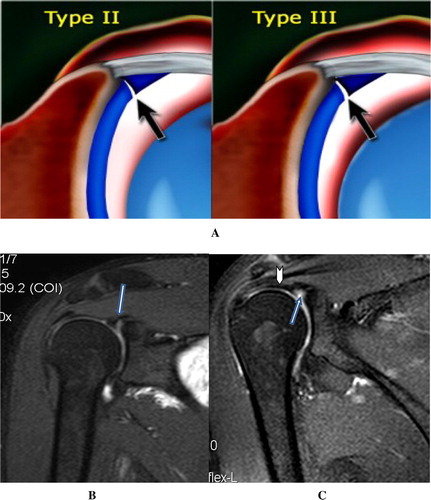
Figure 7 (A, B) Axial MR arthrogram T1 fat sat. Images showing interrupted glenoid attachment of the middle glenohumeral ligament with frayed distorted fibers (arrows).