
Figures & data
Table 1 Age and gender of the patients.
Table 2 The clinical presentation of the patients.
Table 3 Operative data of the two groups.
Table 4 Laminectomy levels and fixation in group I.
Table 5 Instrument profile.
Table 6 Complications in the 2 groups.
Figure 1 (A) Preoperative plain X-ray cervical spine lateral view of a male patient 57 years old presented with quadromyelopathy, operated with cervical laminectomy with lateral mass fixation, (B) Preoperative multislice CT cervical spine, (C, D) Sagittal MRI T2 weighted image of the cervical spine of the same patient before surgery, (E, F) fluoroscopic photo of the cervical spine after the insertion of the screws in lateral view of the same patient, (G) operative photo of the same patient after fixation.
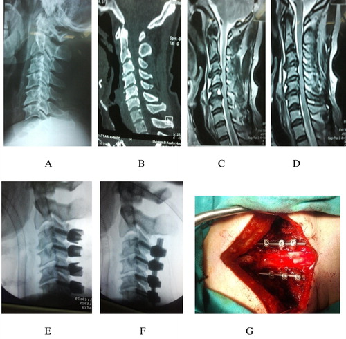
Table 7 Outcome as regards the clinical presentation of the patients.