Figures & data
Table 1 Characteristics of 61 patients with thyroid nodules.
Figure 1 37 year old female presented with papillary thyroid cancer: Coronal T1: shows large heterogeneous nodule mainly involving the right lobe, nodule shows multiple hyper intense foci that denote …hemorrhagic foci, encroached upon the air column. Axial T1 shows ill heterogeneous nodule mainly involving the right lobe. It shows restricted diffusion.
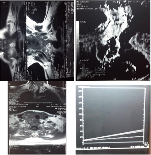
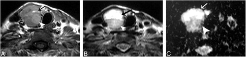
Figure 2 A female patient aged 46 years presented with follicular adenoma. (A) and (B), axial T1- and T2-weighted MR images, respectively, showing a well-defined solitary nodule affecting the right thyroid lobe. (C) An ADC map image with marked hyperintensity of the anterior cystic portion of the nodule (arrow), denotes increased diffusion with a measured ADC value of 2.25 ± 0.18 × 10⁻³ mm2/s and a relatively hypointense posterior solid portion, denotes relatively restricted diffusion with a measured ADC value of 1.2 ± 0.08 × 10⁻³ mm2/s.