Figures & data
Figure 1 Patients with false negative FAST.
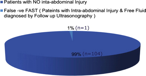
Figure 2 Patients with false positive FAST.
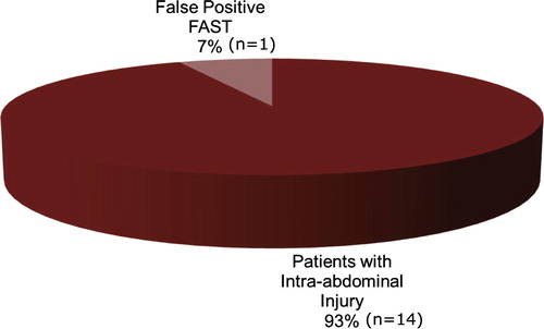
Figure 3 Patients with blunt abdominal trauma.
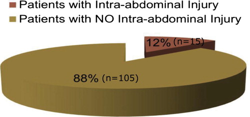
Figure 4 Types of intraabdominal injuries.
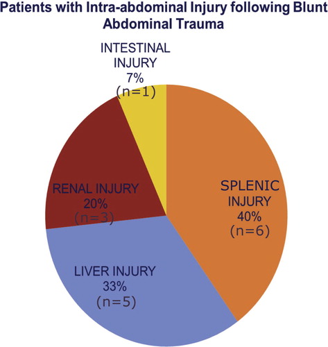
Figure 5 Male patient 26 years old presented to the ER with blunt abdominal trauma following fight. FAST examination performed at the time of presentation showed; (a) Upper pole of the spleen iso to hypoechoic area measuring 1 cm in its maximum diameter (hematoma) and minimal free fluid at the splenorenal angle. (b) Minimal pelvic fluid collection. FAST examination performed after 12 h showed. (c) Upper pole of the spleen shows hypoechoic area 1 cm in its maximum diameter and minimal free fluid at the splenorenal angle. (d) No pelvic fluid collection. Findings of Splenic injury and disappearance of pelvic free fluid suggest stop of bleeding. Management was conservative.
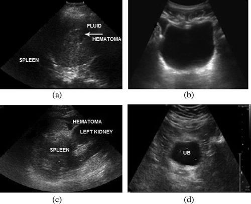
Figure 6 Male patient 25 years old presented to the ER with blunt abdominal trauma following car accident. FAST examination performed at the time of examination. (a) Right lobe of the liver subcapsular area of heterogenous echogenicity measuring 3 × 1 cm in maximum diameters (hematoma), perihepatic moderate free fluid. (b) Marked pelvic free fluid mainly at the right iliac fossa. FAST examination performed after 12 h. (c) The right lobe of the liver subcapsular hypoechoic area measuring 3 × 1 cm in maximum diameter and increased perihepatic free fluid. (d) Increased amount of pelvic free fluid. Findings of liver injury and increased intra-abdominal and intrapelvic fluid (suggesting active bleeding). Management: Surgical treatment.
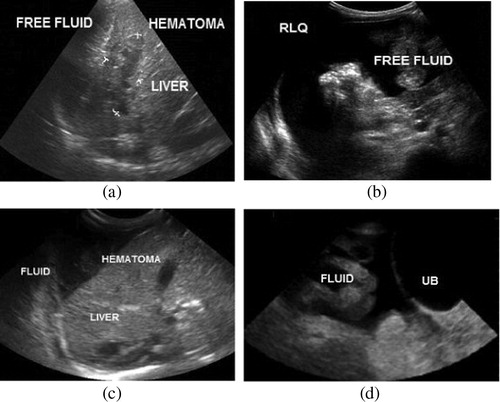
Figure 7 Male patient 55 years old presented to the ER with blunt abdominal trauma following Car accident. FAST examination performed at the time of presentation. (a) The right kidney showed hyperechoic area related to its anterior surface (mostly subcapsular) measuring 3 cm x 1 cm in its maximum diameter not reaching the renal medulla, minimal free fluid at the hepatorenal angle. (b) Minimal pelvic free fluid. FAST examination performed after 12 h. (c) The right kidney showed subcapsular hematoma of heterogenous echogenicity (less echogenic than in FAST) not reaching the renal medulla, decreased amount of free fluid at the hepatorenal angle. (d) Minimal pelvic free fluid. Findings of Right Renal injury and Decreased intra-abdominal and intrapelvic fluid (bleeding). Management: CECT, Conservative treatment.
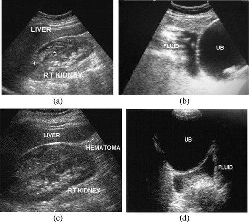
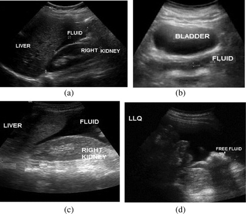
Figure 8 Male patient 35 years old presented to the ER with blunt abdominal trauma following car accident. FAST examination performed at the time of presentation. (a) Minimal free fluid at the hepatorenal angle, and (b) minimal pelvic free fluid. Findings of Positive FAST; however, no gross organ injury is detected. FAST examination performed after 12 h. (c) Increased free fluid at the hepatorenal area. (d) Increased pelvic free fluid. Findings suggestive of active intra-abdominal bleeding and organ injury; however, no gross organ injury is detected. Management: CECT abdomen and pelvis were performed just after FAST examination, and CECT revealed intestinal injury. Sigmoid colostomy was done.