Figures & data
Figure 2 Results of nested PCR of clinical specimens from patients with onychomycosis. Lane M 100-bp DNA ladder (molecular mass markers); lane 1,2,3, nested-PCR-positive specimens (288 bp); lane 4,5 nested-PCR-negative specimens.
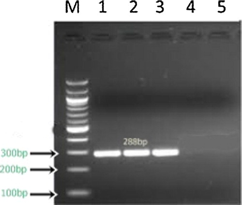
Figure 3 Melting curve analysis of PCR product of pan dermatophyte gene (melts at 81.9 °C).
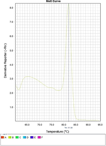
Figure 1 (a) Slide culture of T. mentagrophytes. (b) Culture of T. mentagrophytes on SDA (microscopic). (c) Slide culture of T. verrucosum. (d) Culture of T. verrucosum on SDA. (e) Slide culture of T. rubrum. (f) Culture of T. rubrum on SDA.
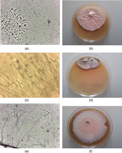
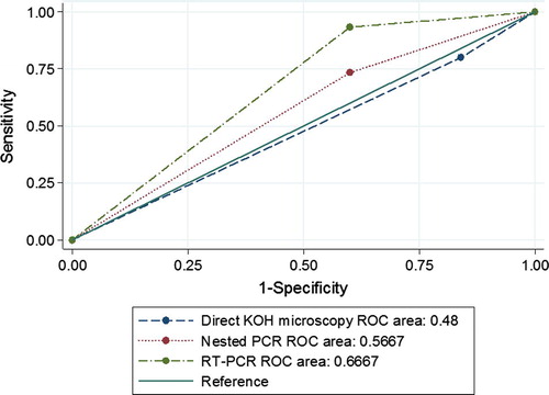
Figure 4 Receiver operating characteristic (ROC) curve for direct KOH microscopy, nested PCR and RT-PCR against the gold standard test (fungal culture) for the diagnosis of onychomycosis (N = 80). KOH microscopy: n = 66 positive and n = 14 negative; Area Under the Curve (AUC) = 0.48, P < 0.001 Nested PCR: n = 52 positive and n = 28 negative; AUC = 0.57, P < 0.001. RT-PCR: n = 58 positive and n = 22 negative; AUC = 0.67, P < 0.001.