
Figures & data
Figure 1 Preoperative CT scan of the patient contrast enhanced CT scan axial view of the neck showing the chondrosarcoma of the hyoid bone on the left side with heterogeneous consistency and peripheral calcification (A), (B) axial view and (C) coronal view.
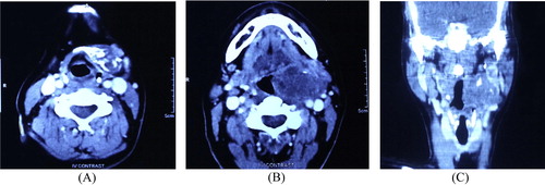
Figure 2 Operative demonstration of the tumor (A) showing the mass attached to the left side of hyoid bone deep to the sternomastoid muscle and (B) the surgical specimen.
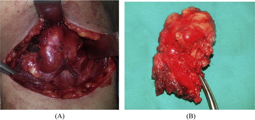
Figure 3 Histopathological examination of the tumor (A) part of a nodule of malignant cartilaginous growth made up of crowded hyperchromatic chondrocytes lying on a chondroid matrix. H&E 200×. (B) Closer view of the hyperchromatic multiple chondrocytes residing in their lacunae and lying on a basophilic chondroid matrix. H&E 300×.
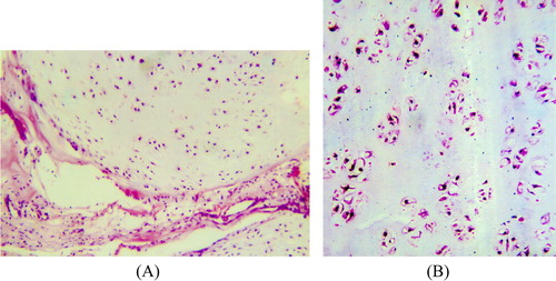
Figure 4 Postoperative imaging contrast enhanced CT scan axial view showing left parapharyngeal metastatic lymph node (white arrow).
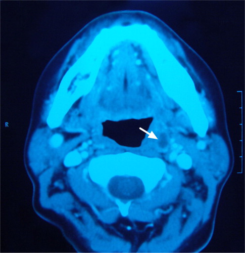
Figure 5 Histopathology of metastatic lymph node (A) showing deposits of chondroid tissue with chondrocytes showing nuclear pleomorphism and mild hyperchromatesia in chondroid matrix H&E 300× (B) with higher magnification H&E 400×.
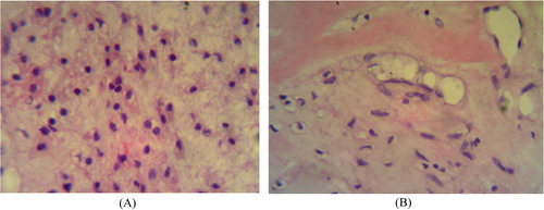
Table 1 Histopathological grades of chondrosarcoma.