
Figures & data
Table 2 Distribution of Patients according to elastogram score in correlation with post operative histopathology.
Figure 1 Rago elastography criteria for assessment of thyroid nodules: (a) score of 1, indicated even elasticity along the entire nodules, (b) score of 2, indicated elasticity along most of the nodule, (c) score of 3 indicated preserved elasticity at the periphery of the nodule, (d) score of 4 indicated no elasticity along the entire nodule, (e) score of 5 indicated loss of elasticity in the whole nodule and along the area showing posterior shadowing. Rago et al. Citation15
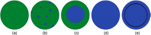
Figure 2 54 years old male patient presented with painless neck swelling since 3 years (A) Transverse grayscale US of a thyroid nodule at the junction between the isthmus and the left lobe showing mainly solid nodule, hypo-echoic to thyroid parenchyma with thin regular halo surrounding the nodule (black arrow). (B) US elastogram showing elasticity score 2 and strain ratio was 1.9. FNAC revealed Bethesda II. Total thyroidectomy was done, and final diagnosis was benign hyperplastic nodule.
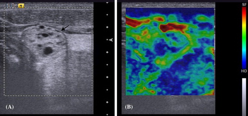
Figure 3 40 years old female patient presented by painless neck lump since two years.(A) Transverse grayscale US of a right thyroid nodule showing a well defined, solid, homogenously slightly hypoechoic nodule (black arrow). (B) US elastogram showing elasticity score 3, strain ratio was 2.4. FNAC was Bethesda IV. Total thyroidectomy was done, and the diagnosis was follicular adenoma.
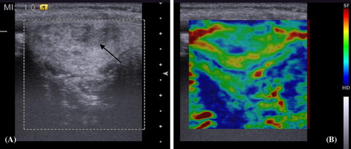
Figure 4 50 years old male patient presented painless neck swelling since one year with rapid growth in the last four months. (A) Transverse grayscale US of the left thyroid lobe showing hypoechoic echotexture and multiple internal echogenic septae (black arrow). (B) US elastogram showing elasticity score 3 and strain ratio 2.1. FNAC showed Bethesda V. Total thyroidectomy was done and the diagnosis was nodular Hashimoto thyroiditis.
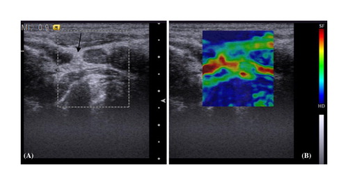
Figure 5 35 years old male patient presented with painless neck lump since one year. (A) Transverse grayscale US of a right thyroid nodule showing predominantly solid nodule, isoechoic, well defined and surrounded by thin regular halo with punctuate foci of micro-calcification (black arrow). (B) US elastogram showing elasticity score 4 and strain ratio 2.7. FNAC revealed Bethesda IV. Total thyroidectomy was done and the diagnosis was follicular carcinoma.

Figure 6 21 years old female patient presented with painless neck swelling since three months. Thyroid function was normal. (A) Transverse grayscale US of a solid nodule at the junction between the isthmus and right lobe showing solid isoechoic echotexture with irregular margin and multiple punctuate echogenic foci of micro calcification (black arrow). (B) US elastogram showing elasticity score 5 and strain ratio was 4.3. FNAC showed Bethesda IV. Total thyroidectomy with neck dissection was done and the diagnosis was papillary carcinoma with nodal metastasis.
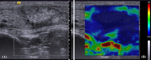
Figure 7 32 years old male patient presented with painless neck swelling since one year. The patient had positive family history of medullary carcinoma, bilateral pheochromocytoma and was diagnosed as MEN IIA. (A) Longitudinal grayscale US of a right thyroid nodule showing hypo-echogenicity, lobulated margin and multiple echogenic foci of micro-calcification (thick white arrow). (B) US elastogram showing elasticity score 5 and strain ratio 4.2. FNAC revealed Bethesda V. Total thyroidectomy was done and the final diagnosis was medullary carcinoma.

Table 1 Final histopathological diagnosis of the examined thyroid nodules.