Figures & data
Figure 1 A photomicrograph of a coronal section in control adult rat cerebral hemisphere showing the location of the entorhinal cortex which is bordered medially by the parasubiculum (P) and laterally by the perirhinal cortex (R). Two main subdivisions of the entorhinal cortex were detected; the lateral (arrow) and the medial (arrow head) entorhinal areas. Curved arrow points to the rhinal fissure (RF) [Gallocyanine stain ×40].
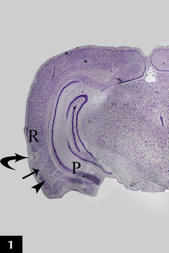
Plate 1 Photomicrographs of a coronal section in the lateral entorhinal area of control group (Figs. 2a, 2b, 2c and 2d). Fig. 2a shows neuronal cells organized in layers I, II, III, IV, V and VI. Ld points to lamina dissecans and RF points to the rhinal fissure. [Gallocyanine stain ×100]. Fig. 2b shows layer I (poorly cellular), layer II (contains cell islands of rounded cells), their nuclei have prominent nucleoli (arrow) and layer III (contains medium pyramidal cells) [Gallocyanine stain ×400]. Fig. 2c shows layer II (contains cell islands of rounded cells), layer III (contains medium pyramidal cells), prominent nucleoli in their nuclei (arrow) and layer IV. (Ld) points to lamina dissecans [Gallocyanine stain ×400]. Fig. 2d shows layer V [contains mainly large pyramidal cells (arrow)] and layer VI contains cells of various sizes and shapes (arrow heads) [Gallocyanine stain ×400].
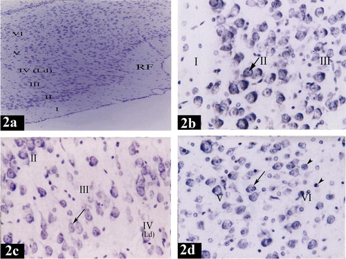
Plate 3 Photomicrographs of a coronal section in the medial entorhinal area of control group (Figs. 3a, 3b, 3c and 3d). Fig. 3a shows neuronal cells organized in layers I, II, III, IV, V and VI. (Ld) points to lamina dissecans [Gallocyanine stain × 100]. Fig. 3b shows layer I (poorly cellular) and layer II (contains cell islands of rounded cells). (Arrows) point to the prominent nucleoli in the nuclei [Gallocyanine stain ×400]. Fig. 3c shows layer III [contains medium pyramidal cells (arrow head)], layer IV, (Ld) points to lamina dissecans and layer V [contains mainly large pyramidal cells (arrow)] [Gallocyanine stain ×400]. Fig. 3d shows layer V [contains mainly large pyramidal cells (arrow)] and layer VI [contains cells of various sizes and shapes (arrow heads)] [Gallocyanine stain ×400].
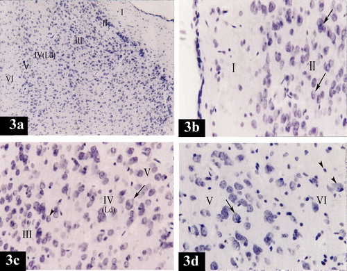
Plate 5 Photomicrographs of a coronal section in the lateral entorhinal area of control group (Figs. 4a and 4b) and treated group (Figs. 4c and 4d). Fig. 4a shows layers I (poorly cellular) and II (contains cell islands of rounded cells). Notice the prominent nucleoli (arrow head) in the nuclei (arrow) [Toluidine blue ×400]. Fig. 4b shows layer III contains medium pyramidal cells (crossed arrow). Their nuclei (arrow) have prominent nucleoli (arrow heads) [Toluidine blue ×400]. Fig. 4c shows dark neurons with hyperbasophilia (arrow heads), diffuse chromatolysis of nuclear chromatin and absence of nucleoli (crossed arrows), absence of nuclei (arrow), degenerative vacuolization (crosses) and intercellular edema (stars) in layers I and II [Toluidine blue ×400]. Fig. 4d shows dark neurons with hyperbasophilia (arrow head), absence of nuclei (arrow) and intercellular edema (star) in layer III [Toluidine blue ×400].
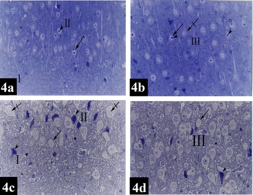
Plate 6 Photomicrographs of a coronal section in the medial entorhinal area of control group (Figs. 4e and 4f) and treated group (Figs. 4g and 4h). Fig. 4e shows layers I (poorly cellular) and II [contains mainly cell islands of rounded cells (arrow head)]. Notice the presence of prominent nucleoli (crossed arrow). Few pyramidal cells (arrow) can be detected within layer II [Toluidine blue ×400]. Fig. 4f shows layer III [contains medium pyramidal cells (arrows) and their nuclei (crossed arrow) with prominent nucleoli (arrow head)] [Toluidine blue ×400]. Fig. 4g shows apoptotic cells (arrowheads), diffuse chromatolysis of nuclear chromatin and absence of nucleoli (crossed arrow), absence of nuclei (arrow) and intercellular edema (star) in layer II [Toluidine blue ×400]. Fig. 4h shows absence of nuclei (arrow), intercellular edema (star) and degenerative vacuolization (arrow head) in layer III [Toluidine blue ×400].
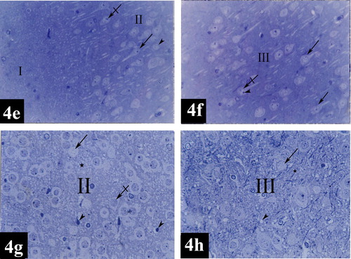
Plate 2 Photomicrographs of a coronal section in the lateral entorhinal area of treated group (Figs. 2e, 2f, 2g and 2h). Fig. 2e shows neuronal cells disorganization in layers I, II, III, IV, V and VI. (Ld) points to lamina dissecans and (RF) points to the rhinal fissure [Gallocyanine stain ×100]. Fig. 2f shows degenerative vacuolization (arrow heads) and intercellular edema (stars) in layers I and II. Layer II shows; apoptotic cells (crossed arrow) characterized by neuronal shrinkage and chromatin condensation, diffuse chromatolysis of nuclear chromatin and absence of nucleoli (arrow) [Gallocyanine stain ×400]. Fig. 2g shows apoptotic cells (crossed arrow), diffuse chromatolysis of nuclear chromatin and absence of nucleoli (arrow) and intercellular edema (star) in layers III and IV. Multinuclear cells are noticed in layer IV (arrowheads). Note a dilated congested blood capillary (Cap) in layer III. (Ld) points to lamina dissecans [Gallocyanine stain ×400]. Fig. 2h shows apoptotic cells (crossed arrow), extensive degenerative vacuolization (arrow heads), diffuse chromatolysis of nuclear chromatin and absence of nucleoli (arrow) and intercellular edema (star) in layers V and VI [Gallocyanine stain ×400].
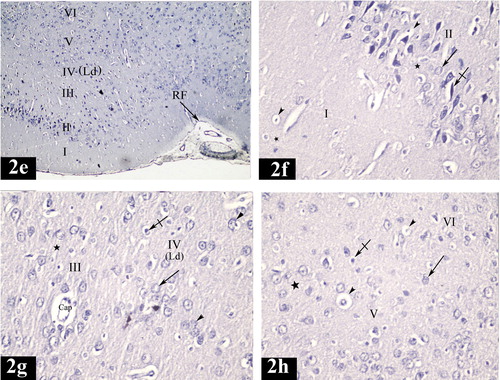
Plate 4 Photomicrographs of a coronal section in the medial entorhinal area of treated group (Figs. 3e, 3f, 3g and 3h). Fig. 3e shows neuronal cells disorganization in layers I, II, III, IV, V and VI. (Ld) points to lamina dissecans [Gallocyanine stain ×100]. Fig. 3f shows degenerative vacuolization (arrow heads), intercellular edema (stars) and apoptotic cells (crossed arrows) in layers I and II. Diffuse chromatolysis of nuclear chromatin and absence of nucleoli (arrow) could be noticed in layer II [Gallocyanine stain ×400]. Fig. 3g shows degenerative vacuolization (arrowhead), intercellular edema (star), apoptotic cells (crossed arrow), diffuse chromatolysis of nuclear chromatin and absence of nucleoli (arrow) in layers III and IV. (Ld) points to lamina dissecans [Gallocyanine stain × 400]. Fig. 3h shows apoptotic cells (crossed arrow), diffuse chromatolysis of nuclear chromatin and absence of nucleoli (arrow), degenerative vacuolization (arrow head) and intercellular edema (star) in layers V and VI [Gallocyanine stain ×400].
Plate 7 Electron micrographs of lateral entorhinal area of control group (Figs. 5a and 5b) and treated group (Figs. 5c and 5d). Fig. 5a shows a granular cell in layer II. The cell has rounded nucleus (N) with fine granular chromatin. The cytoplasm is rich with rough endoplasmic reticulum cisternae (rER), numerous mitochondria (M) and lysosomes (L). Notice the presence of nerve fibers surrounded with myelin sheaths (S) [×5800]. Fig. 5b shows a pyramidal cell in layer III. It has oval nucleus (N) with electron dense heterochromatin. The cytoplasm contains rough endoplasmic reticulum cisternae (rER), mitochondria (M), numerous free ribosomes (R), lysosomes (L) and few vacuoles (V) [×5800]. Fig. 5c shows a granular cell in layer II. The cell shows shrinkage rounded nucleus (N) and chromatin condensation; margination, and clumping with extensive electron dense heterochromatin. The cytoplasm contains free ribosomes (R), distorted rough endoplasmic reticulum cisternae (rER), many degenerative vacuoles (V), lysosomes (L) and disorganized mitochondria (M) [×5800]. Fig. 5d shows a pyramidal cell in layer III. The cell has oval nucleus (N), chromatin condensation; margination and clumping with electron dense heterochromatin. (Arrow) points to irregularity of nuclear envelope. The cytoplasm contains degenerative vacuoles (V), lysosomes (L), free ribosomes (R), mitochondria swollen (M) and distorted rough endoplasmic reticulum cisternae (rER) [×5800].
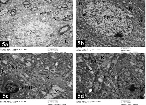
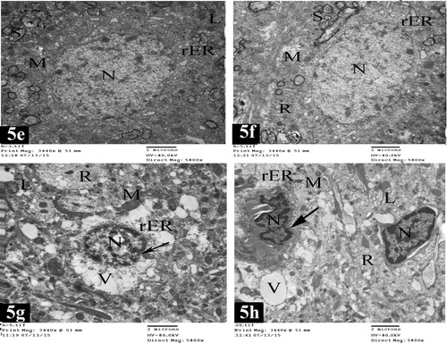
Plate 8 Electron micrographs of medial entorhinal area of control group (Figs. 5e and 5f) and treated group (Figs. 5g and 5h). Fig. 5e shows a granular cell in layer II. The cell contains rounded nucleus (N) with electron dense heterochromatin. The cytoplasm contains rough endoplasmic reticulum cisternae (rER), mitochondria (M) and lysosomes (L). Notice the presence of nerve fibers surrounded with myelin sheaths (S) [×5800]. Fig. 5f shows a pyramidal cell in layer III with oval nucleus (N) and fine granular chromatin. The cytoplasm contains rough endoplasmic reticulum cisternae (rER), mitochondria (M) and free ribosomes (R). Nerve fibers surrounded with myelin sheaths (S) are present [×5800]. Fig. 5g shows a granular cell in layer II. It has shrinkage rounded nucleus (N) and chromatin condensation; margination and clumping with electron dense heterochromatin. (Arrow) points to irregularity of nuclear envelope. The cytoplasm contains numerous degenerative vacuoles (V), lysosomes (L), free ribosomes (R), mitochondria swollen (M) and distorted rough endoplasmic reticulum cisternae (rER) [×5800]. Fig. 5h shows pyramidal cells in layer III. The cells have shrinkage oval nuclei (N) and chromatin condensation; margination and clumping with electron dense heterochromatin. (Arrow) points to marked irregularity of nuclear envelope. The cytoplasm shows numerous degenerative vacuoles (V), lysosomes (L), free ribosomes (R), damaged mitochondria (M) and distorted rough endoplasmic reticulum cisternae (rER) [×5800].
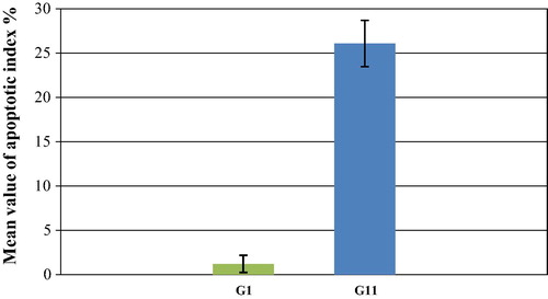
Histogram 1 Mean values ± SD (standard deviation) of apoptotic index % of control (GI) and treated (GII) groups. P ⩽ 0.001 versus control (highly significant).