Figures & data
Figure 1 (A) Anatomic graph of PLC structures, (5) (B) 3D rendering of PLC with the biceps removed demonstrates the Y-shaped arcuate ligament composed of the medial (blue) and lateral (red) limbs attached (green) to the fibular styloid process. FCL, FFL, PFL, and PM are shown.
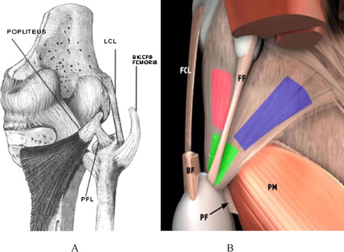
Figure 2 MRI anatomy of knee PLC: (A) Coronal T2-weighted image shows the origin of the lateral gastrocnemius head (arrowhead), femoral attachment of the FCL black arrow), and insertion of the popliteus tendon (white arrow). (B) Sagittal T2-weighted image shows the popliteus tendon (arrow) adjacent to the PHLM (arrowhead) with synovial fluid between them as the popliteus tendon passes through the popliteal hiatus, (C) within popliteomeniscal fascicles (arrow). (D) Axial T2w image: The popliteus tendon (long arrow) posterior to the PHLM, with FCL superficial to it (short arrowhead).Citation4
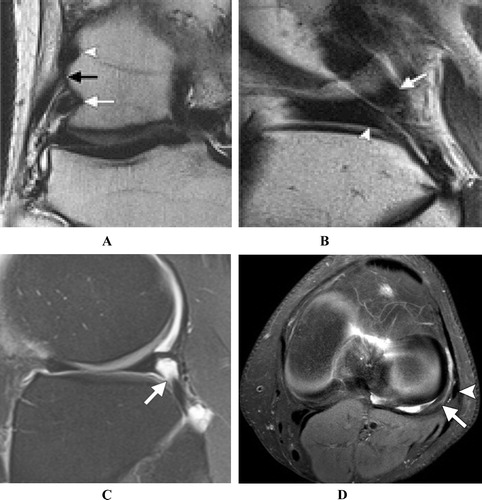
Table 1 Age groups of the patients.
Table 2 Incidence of the different types of PLC structure injuries.
Table 3 Incidence of associated other major compartmental injuries with PLC injury.
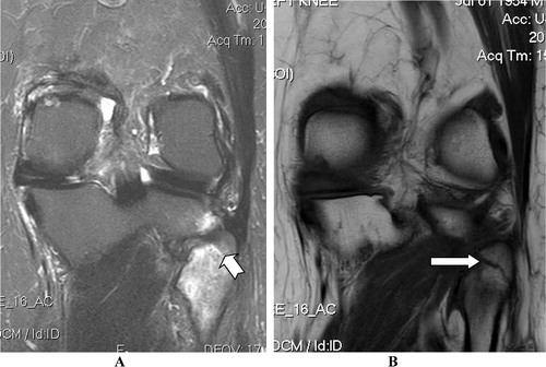
Figure 3 Coronal STIR images showing: (A & B) ITB grade I injury with peri-ligamentous soft tissue edema (Arrow) and intra-articular tibial plateau fracture (Chevron), in association with FCL tear (Curved arrow). (C & D) Another patient with ITB tear (Arrow) associating FCL tear (Chevron).
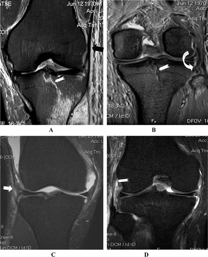
Figure 4 (A & B) Coronal STIR images, (C & D) Axial PD Fat Sat. images, showing aggressive PLC trauma, with avulsed conjoined tendon (biceps and FCL) tear. (E & F) Comparative normal knee: Consecutive axial fat-sat FSE T2 images show normal appearance of the distal bicep tendon joined with FCL to form conjoined tendon, which inserts on lateral aspect of fibular head (Arrows).Citation9
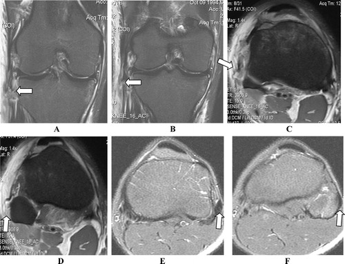
Figure 5 (A & B) Coronal PD-FS (C & D) Sagittal PD-FS images showing torn FCL (Arrow), torn popliteus muscle tendon (Curved arrow), lacerated FPL (Pentagon in A & B) and ACL tear (Notched arrow), together with expected capsular injury seen as extraarticular extravasated synovium (Chevrons).
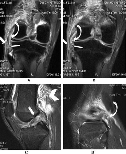
Figure 6 (A–D) Coronal STIR images from posterior to anterior showing partially avulsed popliteal tendon (Arrows) form its femoral attachment, which shows localized marrow edema (Chevrons), with normal FCL (Notched Arrows).
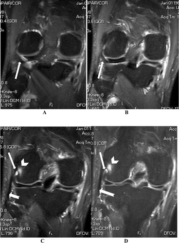
Figure 7 STIR images showing: (A&B) Torn fibular attachment of PFL in RTA patient (Arrows), with intact its popliteus attachment (Curved arrow) and fractured tibial plateau (Notched arrow), in comparison with another normal knee (C & D) which shows intact PFL (Arrows) joining the popliteus tendon (Chevrons).
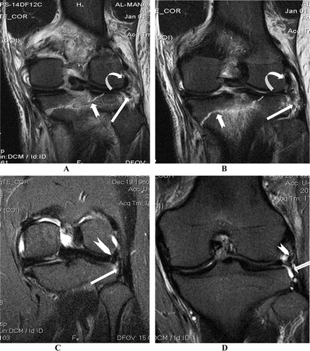
Figure 8 (A & B) axial PD Fat Sat images showing torn arcuate ligament and interrupted PLC joint capsule (Arrows) with posterolateral peri-articular soft tissue edema and lack of joint fluid due to extravasation. (C & D) Another knee showing intact arcuate ligament bordering the joint fluid (Arrows).
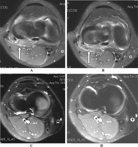
Figure 9 (A) Coronal STIR image showing severe upper fibular marrow edema (Notched arrow). (B) Coronal T1w image of the same patient showing fractured fibular styloid process (Arrow).