Figures & data
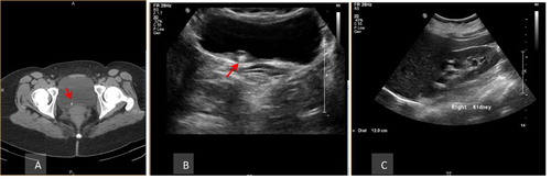
Figure 1 A 45 male patient with left renal colic, (A) axial unenhanced MDCT showing left vesico-ureteral calculus (arrow), (B) Axial prone image, (C & D) unenhanced MDCT with curved multiplanar reformatted coronal and sagittal reconstruction showing the calculus (arrow), (E) Sagittal sonographic image of the left kidney shows moderate hydro-nephrotic and hydro-ureteral changes with the upper ureter measures 8 mm and (F) axial sonographic image of the urinary bladder shows left veiscourteral calculus 6 mm in size (arrow).
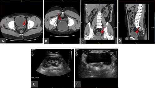
Figure 2 A 41 years old male patient with right renal colic, (A) axial unenhanced MDCT showing right upper ureteral hyper-attenuating calculus with positive rim sign along with moderate hydro-nephrotic and hydro-ureteral changes, (B & C) unenhanced MDCT with curved multiplanar reformatted coronal & sagittal images and (D) sagittal sonographic image of the right kidney shows moderate hydro-nephrotic changes.
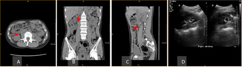
Figure 3 A 37 years old male with right renal colic, (A) axial unenhanced MDCT showing lower calyceal right renal hyper-attenuating calculus (arrow), (B & C) unenhanced MDCT with curved multiplanar reformatted coronal & sagittal images and (D) sagittal sonographic image of the right kidney shows the same finding.
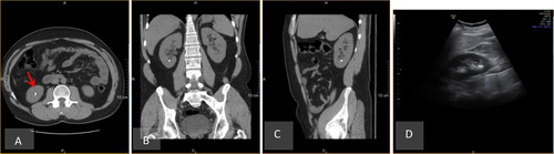
Figure 4 A 42 years old male patient with right renal colic, (A) axial unenhanced MDCT showing right vesico-ureteral calculus (arrow), (B) axial sonographic scan of the urinary bladder shows the calculus at the vesico-ureteral junction with localized hypo-echoic thickening at the bladder wall suggestive of edematous changes (arrow) and (C) sagittal sonographic scan of the right kidney shows moderate hydro-nephrotic changes.