Figures & data
Fig. 1 A 54 years old male with urinary bladder cancer (Invasive TCC, grade III), underwent radical cystectomy and urinary diversion (ileal conduit). Follow up MRU 2 years postoperative was done. (1A) Coronal maximum intensity-projection MR urography images obtained from three-dimensional (3D) T2 turbo spin echo, (1B) coronal T2 WI & (1C) axial T2 WI sequences show a left intra-ureteric defect elicited in conventional MR sequences as soft tissue lesion (arrows) likely meta-synchronous Lesion (proved pathologically to be TCC grade III) with subsequent hydroureter & hydronephrosis .A pelvic cystic fluid collection is also noted.
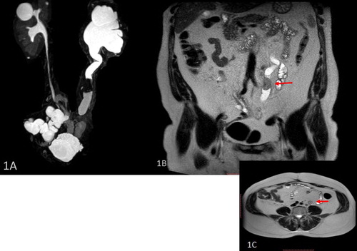
Table 1 Demographic, primary pathology, type of urinary diversion, laboratory features and MRI findings of the patients.
Table 2 MR urography sequences used in Philips Acheiva MR scanner.
Fig. 2 A 61 years old male patient with urinary bladder cancer (Verrucoid SCC, grade I) underwent radical cystectomy, pelvic lymphadenectomy, rectal bladder & colostomy. MRU 1 year postoperative. (2A) Coronal maximum intensity-projection MR urography images obtained from three-dimensional (3D) T2 turbo spin echo & (2B) single shot MR urography image show right uretero-rectal bladder stricture with subsequent hydroureteronephrosis.
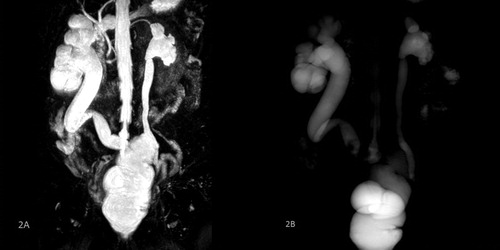
Fig. 3 A 59 years old male patient with cancer bladder (Infiltrating high grade TCC) underwent radical cystectomy, bilateral lymphadenectomy & ileal conduit. MRU is done 6 months postoperative. (3A) Coronal maximum intensity-projection MR urography images obtained from three-dimensional (3D) T2 turbo spin echo, & (3B) Coronal T2 WI show a cystic abdominal fluid collection (lymphocele).
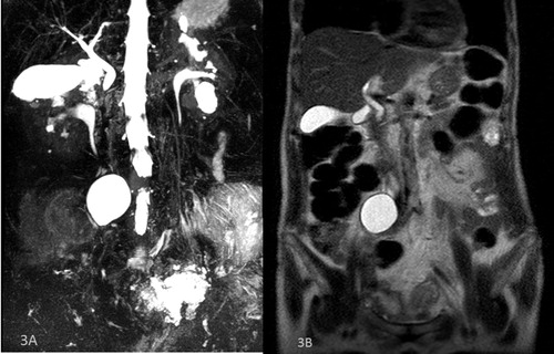
Fig. 4 A 58 years old male patient with cancer bladder (TCC) (Urothelial with squamous differentiation) underwent radical cystectomy, left radical nephrectomy & ileocecal neobladder. MRU 5 years postoperative. (4A) Coronal maximum intensity-projection MR urography images obtained from three-dimensional (3D) T2 turbo spin echo, & (4B) Coronal T2 WI show neobladder mass lesion (recurrence), right upper ureteric stenotic segment and ureteroneobladder stricture.
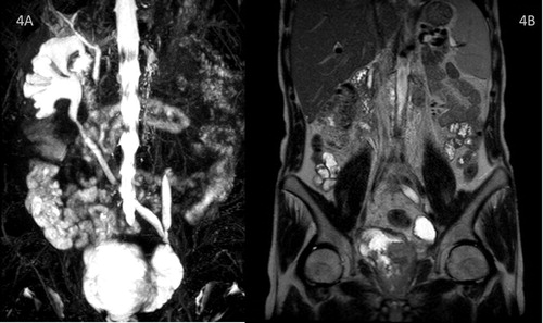
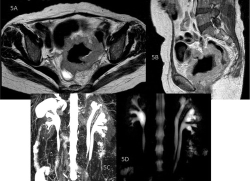
Fig. 5 A 64 years old female with urinary bladder cancer (SCC grade II) underwent anterior pelvic exentration & urinary diversion (Ileal conduit) MRU 1 year postoperative. (5A) Axial T2 WI, (5B) sagittal T1 WI, (5C) Coronal maximum intensity-projection MR urography images obtained from three-dimensional (3D) T2 turbo spin echo & (5D) single shot MR urography image show recurrent pelvic mass with rectal invasion and rectal fistula. Left ureteric complete duplication is noted in the MRU sequences.