Figures & data
Table 1 The clinical presentation and final diagnosis of the examined population. MRS: MR spectroscopy. DTI: Diffusion Tensor Imaging.
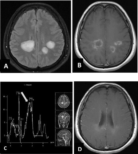
Fig. 1 45 year old male patient with history of left parietal GBM submitted to surgical excision followed by radiotherapy, he presented with this enhancing parenchymal nodule in post contrast axial T1WI (A) that could be recurrence or radiation necrosis. MRS (B) revealed increase in Choline/NAA ratio and Choline/Cr ratio with mild reduced NAA/Cr ratio. (C) DTI revealed decreased FA value with increased ADC value within the enhancing nodule (arrow) denoting high cellularity of the lesion. (D) Is follow up post contrast T1 after 5 months showing progression in size of the lesion which proved to be recurrent Glioblastoma multiform (GBM).
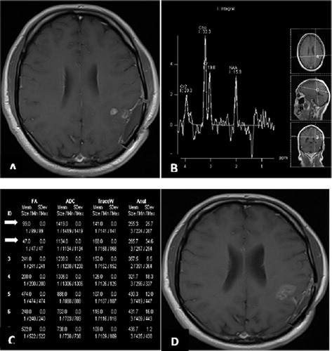
Table 2 Characteristics of MRS in different ring enhancing brain lesions: NAA: N-acetyl aspartate. Cho: Choline. Cr: Creatinine.
Fig. 2 35 year old female patient with multiple patches of deep white matter hyperintensity on axial FLAIR (A), in post contrast axial T1WI (B) these lesions show irregular ring like marginal enhancement, no perifocal edema or mass effect, MR spectroscopy TE 30 from the lesion (C) shows mild elevation of choline/NAA ratio, marked elevation of glutamine peak at 2.1–2.5 ppm (arrow), large lactate peak at 1.3 ppm, and lipid peak at 0.9 ppm, suggesting tumefactive demyelination process. (D) 2 year follow up post contrast axial T1WI after medical treatment of MS shows remarkable regression.