Figures & data
Fig. A1 Cystic, fluctuant and non-tender mass on the right lateral vaginal wall measuring 6 cm by 5 cm.

Fig. A2 Cyst wall excised and sent for histology.

Fig. B1 Low power histologic view of the paravaginal cyst showing predominantly proliferation of numerous small glands of small caliber separated by scanty stroma (H&E ×40 magnification).

Fig. B2 Medium power view of the lesion show proliferation of numerous glands of small caliber (arrow) lined by single to bi-layered cuboidal epithelial cells within its wall that are attenuated in areas. They are separated by intervening scanty stroma. (H & E ×100 magnification).

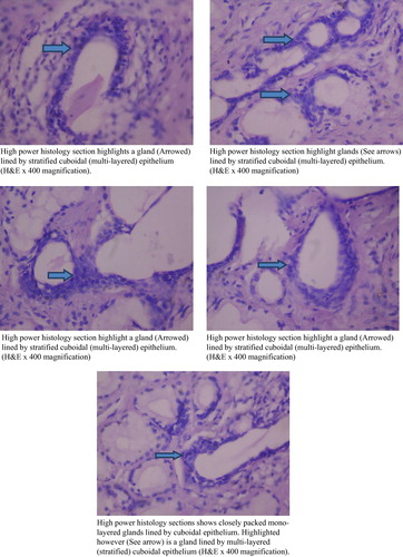
Fig. B3 (a) High power histology section highlights a gland (Arrowed) lined by stratified cuboidal (multi-layered) epithelium (H&E ×400 magnification). (b) High power histology section highlight glands (See arrows) lined by stratified cuboidal (multi-layered) epithelium. (H&E ×400 magnification). (c) High power histology section highlight a gland (Arrowed) lined by stratified cuboidal (multi-layered) epithelium. (H&E ×400 magnification). (d) High power histology sections shows closely packed mono-layered glands lined by cuboidal epithelium. Highlighted however (See arrow) is a gland lined by multi-layered (stratified) cuboidal epithelium (H&E ×400 magnification).