Figures & data
Table 1 The clinical presentations and chief complaints for bladder leiomyoma.
Figure 1 Bladder leiomyoma on pelvic US, showing a smooth endovesical bladder lesion with peripheral hyperechogenicity (yellow arrow).
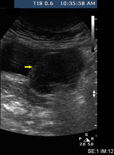
Figure 2 A bladder leiomyoma on CT of the abdomen and pelvis with intravenous contrast medium, showing a well-delineated endovesical bladder tumour, 6 cm × 4.2 cm, arising from the left posterolateral bladder wall.
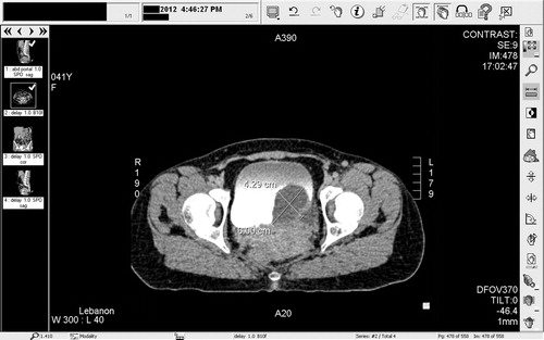
Figure 3 A bladder leiomyoma on CT of the abdomen and pelvis with intravenous contrast medium (coronal view), showing an endovesical bladder tumour, arising from the left lateral bladder wall.
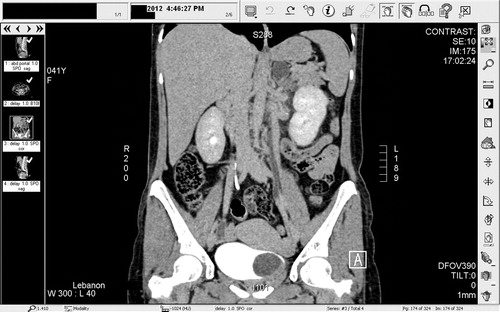
Fig. 4 A bladder leiomyoma on CT of the abdomen and pelvis with intravenous contrast medium (sagittal view), showing an endovesical bladder tumour.
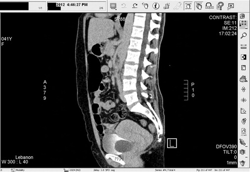
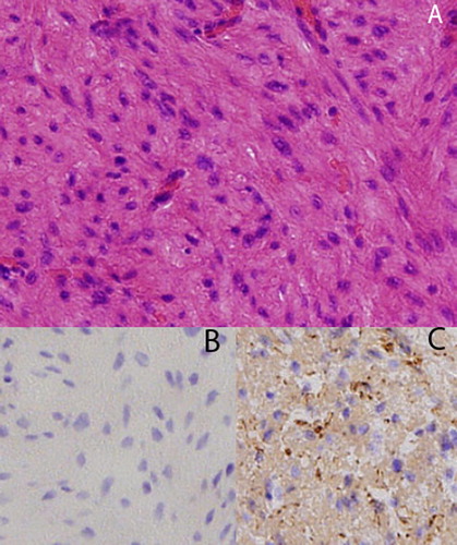
Figure 5 Histopathological studies show that there is a proliferation of spindle-shaped cells, in addition to an eosinophilic cytoplasm and fibres (haematoxylin and eosin, x), with no evidence of mitotic changes or atypia (A). Leiomyomas of the urinary bladder also stain negatively for Ki-67 (B), but they are positive for smooth muscle staining (actin) (C).
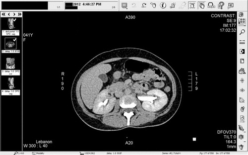
Table 2 Therapeutic options for bladder leiomyoma.
Figure 6 Enhanced CT of the abdomen and pelvis showing left hydronephrosis and a left ‘extra-renal’ pelvis, due to the bladder leiomyoma obstructing the left ureteric orifice.