Figures & data
Figure 1 A diagrammatic depiction of the DIUS technique [Citation8]. (A) A suture is placed between the left colon and the lowest point in the anterior surface of the rectum. (B) An inverted U-shaped sigmoid pouch is detubularised and the posterior wall is closed. Both ureters are re-implanted via the nipple technique [Citation10]. (C) A raw area is created in the posterior rectal and in the descending colon. Both are sutured together. (D) The pouch is closed and the serosal surface of the pouch is connected to the left colon with several interrupted sutures.
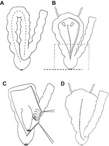
Table 1 Postoperative complications and the changes after DIUS diversion.
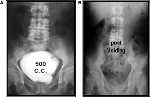
Figure 2 (A) The pouchogram of a patient, showing the good capacity of the pouch. (B) A film taken after emptying in the same patient, showing no residual urine and no reflux to the descending colon.