Figures & data
Table 1 Patient presentation in association to clinical and pathological evidence of BXO.
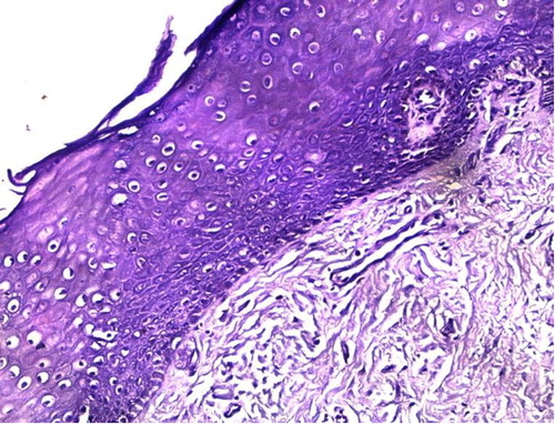
Fig. 1 H&E image showing epidermal atrophy and orthokeratotic hyperkeratosis, showing basal cell vacuolisation, clefting of dermal-epithelial junction; dermis shows dense collagenisation and lymphocytic infiltrate (×200).
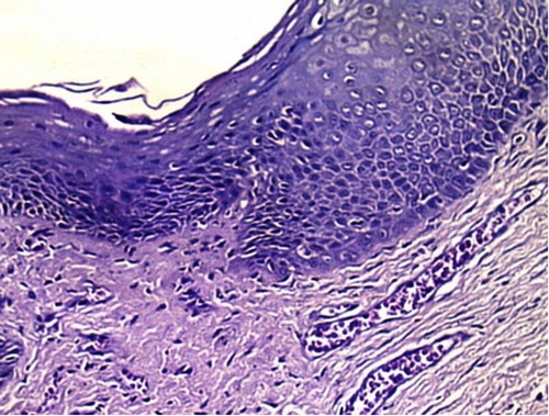
Fig. 2 H&E image showing atrophic epidermis with loss of rete ridges, dermal hyalinisation, thickened basement membrane and marked dermal collagenisation (×200).