Figures & data
Fig. 1 PRISMA flowchart.
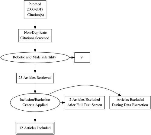
Fig. 2 Operative set-up for robot-assisted microsurgical procedures.
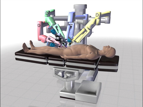
Fig. 3 View from surgeon console during RAVV. Main view from the camera system of the da Vinci robotic platform in the middle, the real-time image from the right side with the andrology optical microscope (×100), and the view from the left side with the VITOM® (Karl Storz GmbH & Co. KG, Tuttlingen, Germany) camera view for enhanced magnification.

Fig. 4 View from surgeon console during RAVE. Main view from the camera system of the da Vinci robotic platform in the middle, the real-time image from the left side with the andrology optical microscope (×100), and the view from the right side with the VITOM camera view for enhanced magnification.
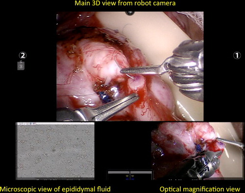
Fig. 5 Intraoperative Doppler US systems (left side audible micro-Doppler, right side visual micro-Doppler).
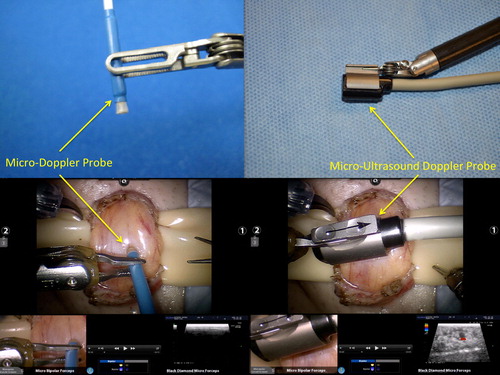
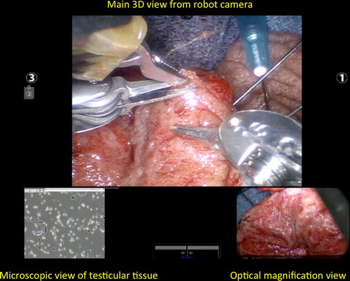
Fig. 6 View from surgeon console during robot-assisted micro-TESE. Main view from robotic camera. Real-time image from andrology laboratory microscope on the left-hand corner. VITOM camera view for enhanced magnification on the right-hand corner.