Liposomal bupivacaine – New trends in Anesthesia and Intensive Care UnitsFootnote
Available online 13 January 2015
Alexandru Florin RogobeteEmergency County Hospital, Clinic of Anesthesia and Intensive Care , Bd. Iosif Bulbuca Nr. 10 , 300736 Timişoara, Romania;“Victor Babeş” University of Medicine and Pharmacy, Faculty of Medicine , Piata E. Murgu 2 , 300041 Timişoara, Romania;West University of Timişoara, Faculty of Chemistry, Biology, Geography , Str. Pestalozzi 16A , 300115Timişoara, Romania
,
Ovidiu Horea BedreagEmergency County Hospital, Clinic of Anesthesia and Intensive Care , Bd. Iosif Bulbuca Nr. 10 , 300736 Timişoara, Romania;“Victor Babeş” University of Medicine and Pharmacy, Faculty of Medicine , Piata E. Murgu 2 , 300041 Timişoara, Romania;[email protected]Correspondence[email protected]
,
Mirela SărăndanEmergency County Hospital, Clinic of Anesthesia and Intensive Care , Department Anesthesia and Intensive Care “Casa Austria” , Bd. Iosif Bulbuca Nr. 10 , 300736 Timişoara, [email protected]
,
Marius PăpuricăEmergency County Hospital, Clinic of Anesthesia and Intensive Care , Bd. Iosif Bulbuca Nr. 10 , 300736 Timişoara, Romania;“Victor Babeş” University of Medicine and Pharmacy, Faculty of Medicine , Piata E. Murgu 2 , 300041 Timişoara, Romania;[email protected]
,
Gabriela PredaWest University of Timişoara, Faculty of Chemistry, Biology, Geography , Str. Pestalozzi 16A , 300115Timişoara, Romania;[email protected]
,
Maria Corina DumbuleuEmergency County Hospital, Clinic of Anesthesia and Intensive Care , Bd. Iosif Bulbuca Nr. 10 , 300736 Timişoara, Romania;[email protected]
,
Corina Vernic“Victor Babeş” University of Medicine and Pharmacy, Faculty of Medicine , Piata E. Murgu 2 , 300041 Timişoara, Romania;[email protected]
,
Emil Robert Stoicescu“Victor Babeş” University of Medicine and Pharmacy, Faculty of Medicine , Piata E. Murgu 2 , 300041 Timişoara, Romania;[email protected]
&
Dorel SăndescEmergency County Hospital, Clinic of Anesthesia and Intensive Care , Bd. Iosif Bulbuca Nr. 10 , 300736 Timişoara, Romania;“Victor Babeş” University of Medicine and Pharmacy, Faculty of Medicine , Piata E. Murgu 2 , 300041 Timişoara, Romania
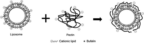
Figure 3 Liposomal bufalin functionalized with citrus pectin [Citation11].
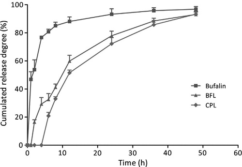
Figure 4 In vitro release kinetic profiles of bufalin from liposomal bufalin (BFL), liposomal bufalin functionalized with pectin citrus (CPL) compared to freely soluble bufalin [Citation11].
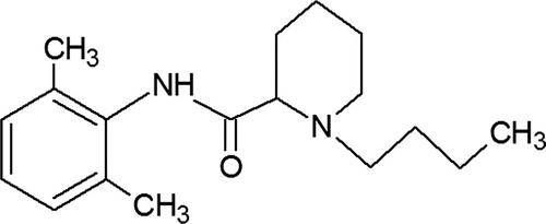
Figure 5 Bupivacaine – chemical structure.
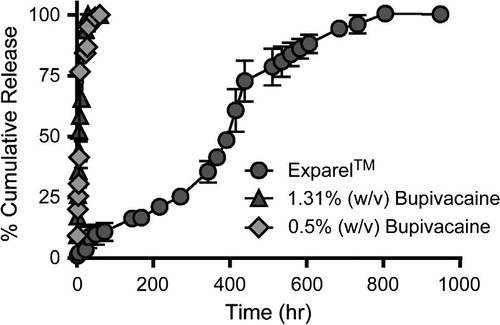
Figure 6 Cumulative release of bupivacaine form Exparel™ and release of unencapsulated 1.31% w/v and 0.5% w/v bupivacaine HCl [Citation57].
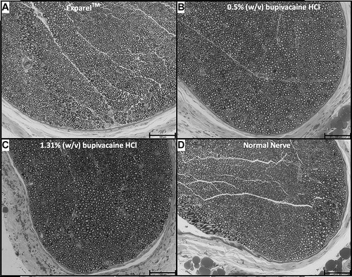
Figure 7 Sciatic nerve samples harvested from rats 4 day after injection with either Exparel™ (A), bupivacaine HCl 0.5% w/v (B), bupivacaine HCl 1.31% w/v (C) or uninjected sciatic nerve (D) [Citation57].
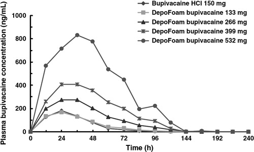
Figure 8 Plasma bupivacaine concentration after administration of DepoFoam bupivacaine or bupivacaine HCl to patients undergoing total knee arthroplasty [Citation62].
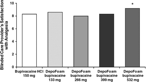
Figure 9 Mean blinded care provider’s satisfaction with analgesia (rating scale: 0 = completely unsatisfied patients analgesia and 10 = completely satisfied patients analgesia) [Citation62].