Figures & data
Table 1 Morphometric analysis of different developing stages of Chameleo chameleon in (mm).
Fig. 1 Lateral view photomacrographs of different stages of developing Chameleo chameleon.
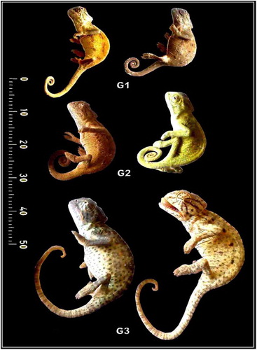
Fig. 2 (A-C): Photomacrographs of lateral and dorsal views of retracted and protruded tongue of Chameleo chameleon. (Abbreviations: Ac, accelerator linguae; CB: ceratobranchial; E, eye; Hg, hyoglossus; LP, lingual pouch; Tp, tongue pad).
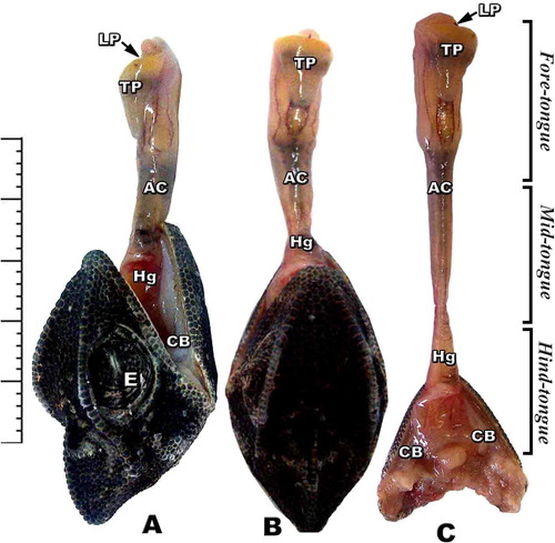
Fig. 3 (A-F): Scanning electron micrograph of dorsal view of tongue of Chameleo chameleon. (Abbreviations: D, Dimple; MS, Muscle strands; LPa; Lingual papillae; LPi, lingual pits; MuS, Mucous secretion; Tp, Tongue pad).
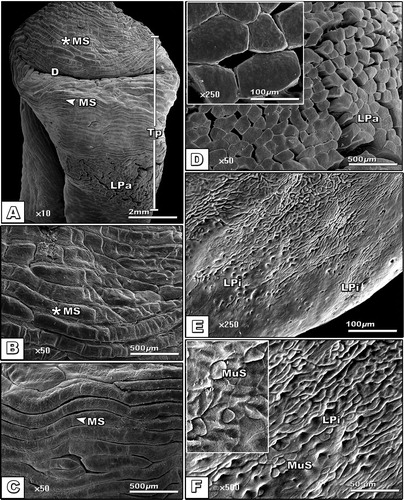
Table 2 Length (μm) and numbers (No./100 μm) of different types of lingual filiform papillae of different developing stages of Chameleo chameleon.
Fig. 4 Mean numbers of various shapes of lingual filiform papillae of different developing stages of Chameleo chameleon. Each result represents the mean ± SE (n = 5) per each 100 μm.
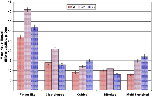
Fig. 5 Mean length (µm) of various shapes of lingual filiform papillae of different developing stages of Chameleo chameleon. Each result represents the mean ± SE (n = 5) per each 100 μm.
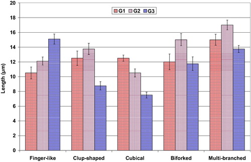
Fig. 6 (A-D): Photomicrograph of histological sections of tongue of Chameleo chameleon. HX-E A. Gross sagittal histological section of tongue pad showing dense distribution of dorsal lingual papillae and lingual glands. B. Sagittal section of medium tongue region. C-D. Transverse histological section of middle cylindrical tongue core showing central cartilaginous compartment ensheathed by connective tissue layer followed by longitudinal and circular muscle layers. (Abbreviations: AcM, Accelerator muscles; Ch, chondrocyte; CILS, central intralingual sheath; CM, circular muscle; CT, connective tissue sheat; EP, enteroglossal process; Epm, epimysium; Hg, hyoglossus muscle; ILS, intralingual sheath; LM, longitudinal muscle; LPa, lingual papillae; MG, mucous gland; PILS, peripheral intralingual sheath; RM, retractor muscle; SM, smooth muscle; Tp, tongue pad).
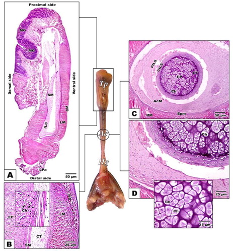
Fig. 7 (A-C1): Photomicrograph of histological sections of tongue of Chameleo chameleon. HX-E (Abbreviations: BF, Biforked; C, clup-shaped; Cu, cubical; DEp, Dorsal epithelium; F, Finger-like; LPa, lingual papillae; MB; Multi-branched MD, mucous duct; MF, muscle fibers; MG, mucous gland).
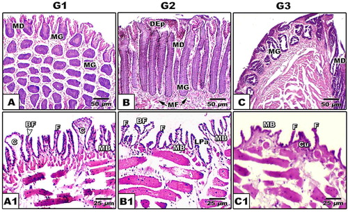
Fig. 8 (A-C2): Photomicrograph of histological sections of tongue of Chameleon chameleon. Alcian-PAS. A-A2. G1 showing alcian positive branched alveolar glands distributed in lamina properia (A), PAS positive alveolar and tubular glands (MG) (A1) & alcian positive alveolar gland (A2). B-B2. G2 Showing negative staining alveolar gland (B) and alcian positive alveolar glands (B1 & B2). C-C2. G3 showing alcian positive branched alveolar gland (C) and PAS positive staining of both tubular and branched alveolar glands (C1 &C2). (Abbreviations: AG, alveolar gland; BAG, branched alveolar gland; DEp; Dorsal epithelium; LP, lamina proporia; MD, mucous duct; MG, mucous gland). (*), Positive PAS staining; The arrow head (
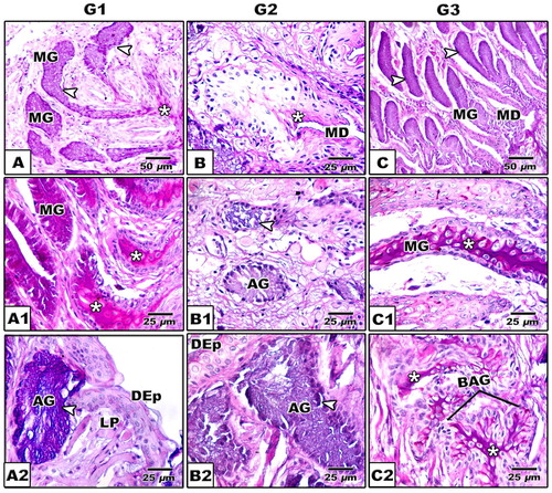
Table 3 Immunoreactivity of CK, SCF and AB/PAS of tongue of G1, G2 and G3 of Chameleo chameleon.
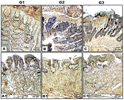
Fig. 9 (A-C1): Photomicrographs of formalin-fixed, paraffin-embedded tongue of Chameleo chameleon, immunohistochemically stained with anti-CK. A-A2. G1. B-B2. G2. C-C2. G3. Note mild cytokeratin reaction in G1 and intense staining in G2 & G3 in lingual mucosa. (Abbreviations; AG, alveolar gland; DEp; Dorsal epithelium; MD, Mucous duct; MG, mucous gland).
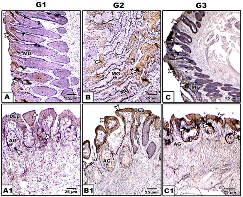
Fig. 10 (A-C1): Photomicrographs of formalin-fixed, paraffin-embedded tongue of Chameleo chameleon, immunohistochemically stained with anti-SCF. A-A2. G1. B-B2. G2. C-C2. G3. Note mild stem cell reaction in G1 and moderate staining in G2 & G3 in both lingual mucosa and lingual glands. (Abbreviations: AG, alveolar gland; BAG, branched alveolar gland; DEp; dorsal epithelium; MD, mucous duct; MG, mucous gland).