Figures & data
Fig. 1 Vincristine loaded folic acid-chitosan conjugated nanoparticles of 4:25 formulation (a) average size and polydispersity index, and, (b) and zeta potential.
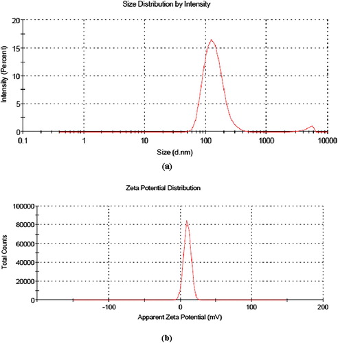
Table 1 Average size, polydispersity index and zeta potential of vincristine loaded folic acid-chitosan conjugated nanoparticles at pH 5.
Table 2 Encapsulation efficiency (%) and loading capacity (%) of vincristine loaded folic acid-chitosan conjugated nanoparticles.
Table 3 Effect of storage on average size, polydispersity index and zeta potential of blank and vincristine loaded folic acid-chitosan conjugated nanoparticles in sodium phosphate buffer (pH 7.4) at 4 °C after 10 days.
Fig. 2 FTIR spectra showing shifting in peaks due to loading of vincristine (a) blank FA-CS NPs and vincristine loaded FA-CS NPs at different formulations, (b) 1:25, (c) 2:25, (d) 3:25, (e) 4:25
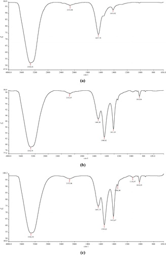
Fig. 3 SEM images of nanoparticles showing spherical shaped (a) blank FA-CS NPs and vincristine loaded FA-CS NPs at different formulations, (b) 1:25, (c) 2:25, (d) 3:25, (e) 4:25.
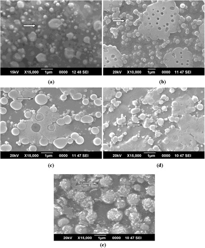
Fig. 4 Transmission electron microphotographs of blank and vincristine loaded FA-CS nanoparticles showing encapsulation of vincristine (arrows) (a) blank and different formulations, (b) 1:25, (c) 2:25, (d) 3:25, (e) 4:25.
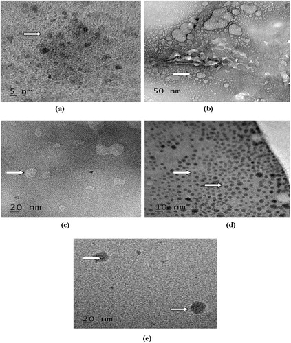
Fig. 5 Cumulative release (%) behavior of vincristine from folic acid-chitosan conjugated nanoparticles for different formulations (1:25, 2:25, 3:25, 4:25) showing slow and sustained release in phosphate buffered saline (pH 6.7).
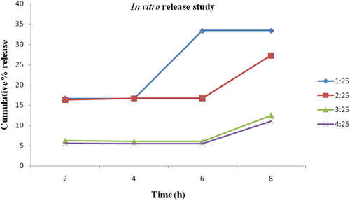
Fig. 6 Cellular uptake of nanoparticles in NCI-H460 cells 40× (a) control, (b) enhanced fluorescence with vincristine loaded folic acid-chitosan conjugated nanoparticles.
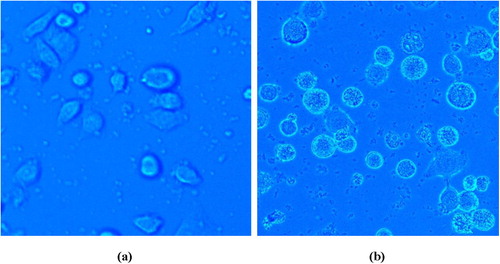
Table 4 Cell viability (%) of blank and vincristine loaded FA-CS nanoparticles at different concentrations.
Fig. 7 Fluorescent micrographs showing effect of nanoparticles in NCI-H460 cells (i) intracellular ROS level, (ii) mitochondrial transmembrane potential, (iii) apoptotic morphological changes.
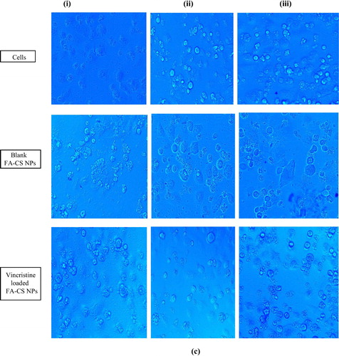
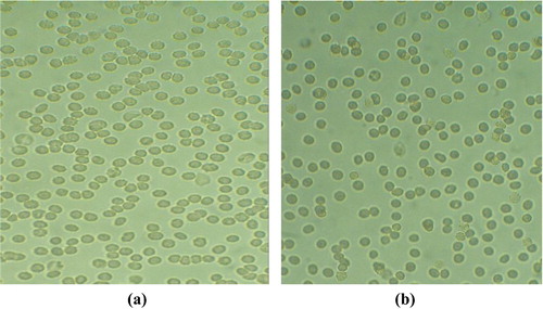
Fig. 8 Light microphotographs of erythrocytes aggregation (a) control, (b) vincristine loaded folic acid-chitosan conjugated nanoparticles.