Figures & data
Fig. 1 The effect of CO extract against PE-induced contraction in endothelium-intact (E+) and endothelium-denuded (E-) aortic rings. Relaxation was calculated as % pre-contraction induced by (0.1 μM) PE. Values are expressed as mean ± S.E.M. where n = 6.
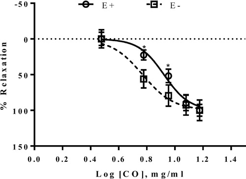
Fig. 2 Relaxant response induced by CO extract in endothelium-intact aortic ring precontracted with (0.1 μM) PE in the presence and absence (control) of L-NAME (100 μM). Values are expressed as mean ± S.E.M. where n = 6.
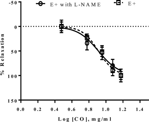
Fig. 3a Inhibitory effect of EC50 of CO on the extracellular Ca2+-induced contraction ranging from 0.3 mM to 10.0 mM in endothelium-denuded aortic ring precontracted by 60 mM KCl in Ca2+-free Krebs solution. Control group was not pre-incubated with CO. Values are expressed as mean ± S.E.M. where n = 4.
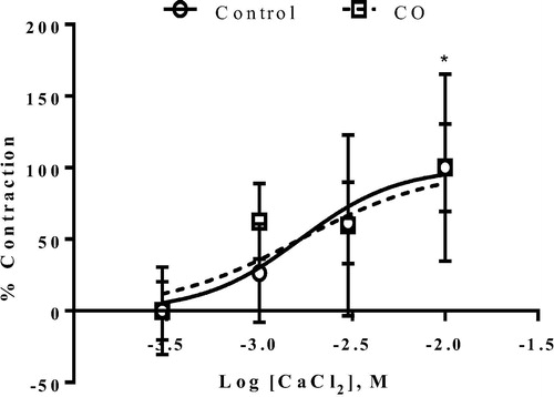
Fig. 3b Inhibitory effect of EC50 of CO at on the extracellular Ca2+-induced contraction at concentration from 0.3 mM to 10.0 mM in endothelium-denuded aortic ring precontracted by (0.1 μM) PE in Ca2+-free Krebs solution. Control group was not pre-incubated with CO. Values are expressed as mean ± S.E.M. where n = 4.
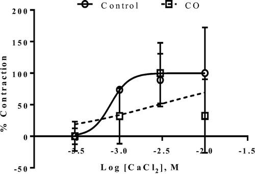
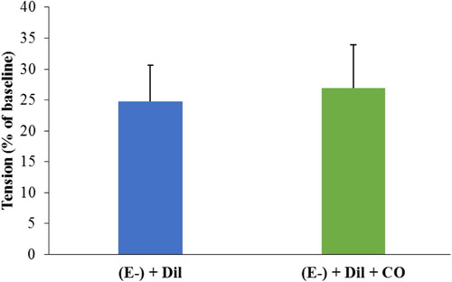
Fig. 4 The effect of CO extract on the intracellular Ca2+ released by the sarcoplasmic reticulum. Endothelium-denuded aortic ring was incubated with diltiazem (1 μM) before addition of (0.1 μM) PE. The rings were then exposed to 4.95 mg/ml of CO extract. The tension produced by the ring was compared before and after the addition of the extract. Values are expressed as mean ± S.E.M. where n = 4.