Figures & data
Table 1 Phytochemical ingredients of aqueous extract of Ocimum gratissimum leaves.
Fig. 1 Effects of AEOGL on percentage weight gain of rats induced colitis by acetic acid. Values are given as Mean ± SEM (n = 5) * = significantly different from control group (p < 0.05); α = significantly different from acetic acid group (AA) (p < 0.05).
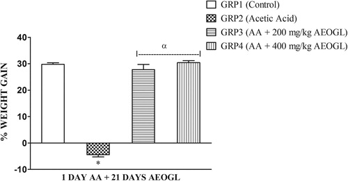
Table 2 Effects of AEOGL on diarrhea score and ulcer parameters of rats induced colitis by acetic acid.
Table 3 Effects of AEOGL on hematological parameters of rats induced colitis by acetic acid.
Fig. 2 Effects of AEOGL on nitric oxide (NO) of rats induced colitis by acetic acid. Values are given as Mean ± SEM (n = 5); * = significantly different from control (p < 0.05); α = significantly different from acetic acid group (AA).
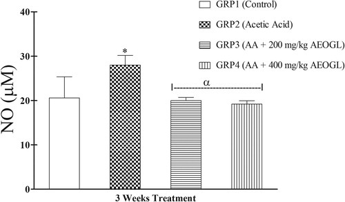
Fig. 3 Effects of AEOGL on myeloperoxidase (MPO) of rats induced colitis by acetic acid. Values are given as Mean ± SEM (n = 5); * = significantly different from control (p < 0.05); α = significantly different from acetic acid group (AA) (p < 0.05).
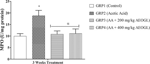
Fig. 4 Effects of AEOGL on superoxide dismutase (SOD) of rats induced colitis by acetic acid. Values are given as Mean ± SEM (n = 5); * = significantly different from control (p < 0.05); α = significantly different from acetic acid group (AA) (p < 0.05).
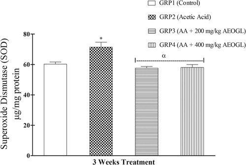
Fig. 5 Effects of AEOGL on reduced glutathione (GSH) of rats induced colitis by acetic acid. Values are given as Mean ± SEM (n = 5); * = significantly different from control (p < 0.05); α = significantly different from acetic acid group (1 day AA) (p < 0.05).
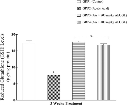
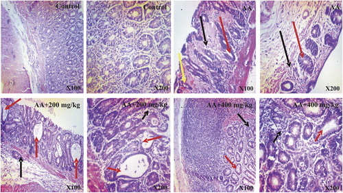
Fig. 6 Photomicrographs of colon tissues showing the effects of AEOGL administration following acetic acid-induced colitis. Photomicrograph of the colon of Control (CN), Acetic Acid (AA), AA + 200 mg/kg and AA + 400 mg/kg. CN shows presence of intact T-lymphocytes and intraepithelial lymphocytes. Abundant or proportionate goblet cells and absorptive cells. In contrast, AA shows severe mucosal atrophy, hyperchromatic nuclei (black arrow), loss of goblet cells, crypts distortions and abscesses (red arrow), hyperemia and tissue hemorrhage (yellow arrow). 200 and 400 mg/kg (Groups 3 and 4) AEOGL treated group showed greater signs of improvement, revealing higher/greater numbers of crypts/glands. They showed high glandular/mucosal height (black arrow), prominent intraepithelial lymphocytes, signs of crypt distortions and abscesses (red arrow). (H & E Stain; ×100 & 400).