Nashwa A. EzzeldeenDepartment of Microbiology, Faculty of Veterinary Medicine, Cairo University, Egypt;Department of Biology, Faculty of Science, Taif University, Kingdom of Saudi Arabia;[email protected] (N.A.Ezzeldeen)
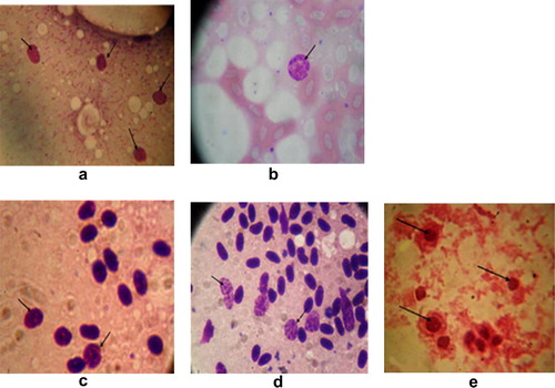
Figure 1 Chlamydial inclusion bodies in the impression smears. (a–d) Chlamydial inclusions in liver, lung, heart and spleen impression smears, respectively, of internal organs of the examined birds stained with Giemsa stain. (e) Chlamydial inclusions in the infected yolk sac membrane stained with Giménez stain.
Table 1 Direct detection of chlamydial inclusion bodies within the examined samples.
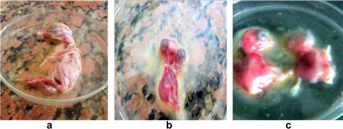
Figure 2 (a) Normal chicken embryo. (b and c) Chicken embryos growth abnormalities: dwarfism and congestion of chicken embryos among the inoculated eggs.
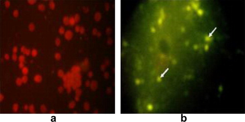
Figure 3 Chlamydial inclusions in the infected yolk sac membrane stained with FA. (a) Negative impression smear for Chlamydia after staining with FA. (b) Positive impression smears for Chlamydia using FA.
Table 2 Detection of Chlamydia psittaci antibodies in the collected serum samples of Hoopoe and Cattle Egret by complement fixation test (CFT).
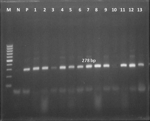
Figure 4 The expected amplified product of 16S rRNA gene specific for family Chlamydiaceae at 278 bp. Lane M: 100 bp DNA ladder (Invitrogen), Lane N: negative control, Lane P: positive control, Lanes 1–9 and 11–13: positive samples and Lane 10: negative sample.
Table 3 Comparison of the percentages of positives yielded by different diagnostic methods used for detection of chlamydiae in Hoopoe and Cattle Egret samples.