746
Views
12
CrossRef citations to date
0
Altmetric
Full Length Articles

Enhanced electroscalpel incisional wound healing potential of honey in wistar ratsFootnote

, , &
Pages 128-134 | Received 14 Aug 2017, Accepted 21 Oct 2017, Published online: 03 May 2019

Figures & data

Table 1 Evaluation of wound exudation, edge edema and hyperemia in electro-scalpel and cold scalpel induced wounds in rats.

Fig. 1 Gross examination of wounds: A – wounds created with electroscalpel at day 5 still having exudation and thick granulation tissue. B – Wounds created with cold scalpel at days 5 already healing progress and contraction of wound surface. C – Wounds created with electroscalpel at day 14 showing wound surface contraction. D – Wounds created with cold scalpel at days 14 already contracted. (S = silver sulphadiazine, C = control, H = honey).

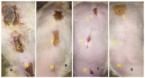
Table 2 Mean daily percentage wound contraction in electroscalpel and cold scalpel induced wounds in rats.

Fig. 2 Photomicrographs of electroscalpel wounds at day 7: A – (SSD) shows moderate granulation tissue (blue arrow). B – (Control) show granulation tissue (blue arrow). C (Honey) shows presence of marked cellular infiltrates (black arrow) and granulation tissue (blue arrow). HE ×400.
Fig. 3 Photomicrographs of coldscapel wound at day 7: A – (SSD) shows severe granulation (blue arrow), and moderate angiogenesis (white arrow). B (Control) shows moderate granulation (blue arrow), and moderate angiogenesis (white arrow). C (Honey) shows epithelialization (green arrow), moderate granulation (blue arrow), mild angiogenesis (thin arrow) and moderate fibrosis. HE ×400.

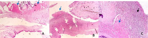
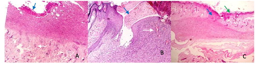
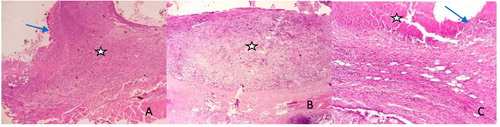
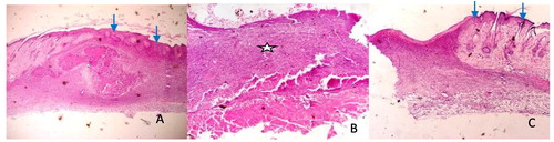
Table 3 Wound histologic indices and inflammatory cell counts of electroscalpel and cold scalpel induced wounds in rats at day 5, 7 and 14.

Fig. 4 Photomicrographs of electroscalpel wounds at day 14: A (SSD) shows granulation (asterick), moderate contraction and epithelialization (blue arrow). B (Control) shows abundant granulation tissue (asterick) with loose connective tissue and no contraction. C (Honey) shows wound contraction (white arrow), moderate granulation (asterick), angiogenesis and epithelialization (blue arrow) HE ×400.
Fig. 5 Photomicrographs of coldscapel wound at day 14: A (SSD) shows complete epithelialization (blue arrow) and wound contraction. B (Control) shows granulation tissues (asterick), and moderate angiogenesis. C (Honey) shows complete epithelialization (blue arrow) and loose connective tissue. HE ×400.