1,948
Views
73
CrossRef citations to date
0
Altmetric
Full Length Articles

Evaluation of bull spermatozoa during and after cryopreservation: Structural and ultrastructural insightsFootnote

, , , &
Pages S49-S56 | Received 08 Sep 2017, Accepted 12 Nov 2017, Published online: 03 May 2019

Figures & data

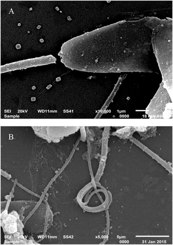
Fig. 1 Representative images of scanning electron microscopy showing examples of sperm cell abnormalities which occur during cryopreservation of bull semen. A: Detached and cracked head, B: coiled tail.

Table 1 The effect of different processing steps of semen cryopreservation on sperm motility, viability and morphological abnormalities examined by light microscopy (mean percentage ± SEM, n = 5).
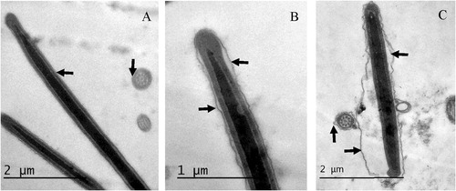
Fig. 2 Representative images of sperm head showing patterns of plasma membrane integrity (arrows) at different stages of semen processing and cryopreservation. A: Intact plasma membrane in fresh semen sample. B: Slightly swollen PM in diluted semen, C: Swollen undulating PM in frozen thawed sample. Cross sections of sperm cells in A and C show the same PM integrity patterns.

Table 2 The effect of different processing steps of semen cryopreservation on sperm plasma membrane (PM) and acrosome reaction (AR) examined by transmission electron microscopy.
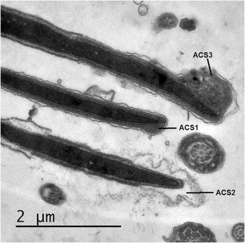
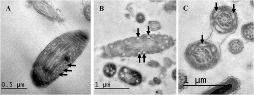
Fig. 3 Representative micrographs showing ultrastructural changes in sperm acrosome. ACS1: Intact acrosome, ACS2: typical acrosome reaction (AR) and ACS3: Atypical AR.
Fig. 4 Mitochondrial morphology and damage induced by cryopreservation (arrows). Longitudinal Ultrathin section of raw semen showing a sperm mid piece. A: normal mitochondria; B and C: damaged mitochondria with distorted cristae. Magnification 10,000×.

Table 3 The effect of different processing steps of semen cryopreservation on sperm mitochondrial damage examined by transmission electron microscopy.
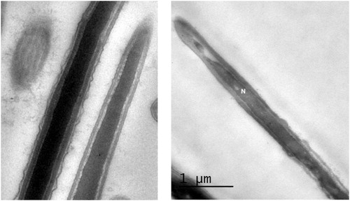
Fig. 5 Nuclear morphology and damage induced by cryopreservation. Longitudinal Ultrathin section of the head piece showing abnormal nucleus (N, right) with big nuclear vacuole containing electron dense material and indicating chromatin damage compared to normal (left) nucleus with homogenous condensed chromatin.

Table 4 The effect of different processing steps of semen cryopreservation on sperm DNA damage examined by transmission electron microscopy, TB staining and SCSA.