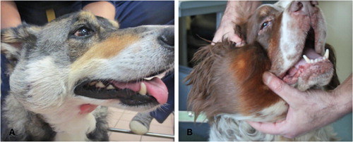
ig. 1 A: Photograph of case 1 showing a well-defined soft tissue mass on the right parotid region. B: Photograph of case 2 showing a right retromandibular soft tissue mass.
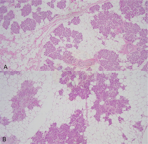
ig. 2 A: Microphotograph of excised tissue from case 1 showing an admixture of mature adipocytes interspersed with well-differentiated salivary acini and few salivary ducts. Haematoxylin and eosin stain, 4×. B: Microphotograph of excised tissue from case 2 showing a similar admixture of adipocytes and well-differentiated salivary acini. Haematoxylin and eosin stain, 4×.
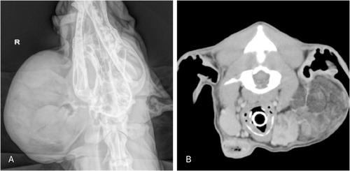
ig. 3 A: Radiographic image of the head of the case 2 showing a soft tissue opacity on the right parotid region and no evidence of osteolyis. B: Transverse plan image of computerized tomography from the head of the case 2 showing a multi-lobulated infiltrative mass occupying the right parotid gland. No evidence of osteolysis was present. The right mandibulary gland had an heterogenous appearance but histology showed normal salivary gland tissue. On the left side, just ventral to the left mandibulary salivary gland there was a soft tissue opacity structure with air-filled, of unknown significance, that had cytological features of inflammation and disappeared with an antibiotic course.
able 1 Description of the cases of fat-containing salivary gland lesions found on a literature review, including original diagnoses and proposed diagnoses in the authors’ opinion.