Chronological analysis of the damages caused by the metacercariae of Centrocestus formosanus in the gills of Cyprinus carpio and lesions caused by the adult flukes in Ardeola ralloides: An experimental studyFootnote
Peer review under responsibility of Faculty of Veterinary Medicine, Cairo University.
Dona H.N. MunasingheDepartment of Zoology, Faculty of Science, University of Ruhuna, Matara, Sri Lanka
&
Appudurai ArulkanthanDepartment of Veterinary Pathobiology, Faculty of Veterinary Medicine and Animal Science, University of Peradeniya, Peradeniya, Sri Lanka
Pages 165-171
|
Received 26 May 2018, Accepted 18 Aug 2018, Published online: 03 May 2019
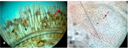
ig. 2 Encysted metacercariae. (a) Wet mounts of the gills of an infected Koi carp with encysted metacercariae (arrow) of C. formosanus (×40). (b) Encysted metacercaria with eye spots (arrow) after 6 days of infection (×100).
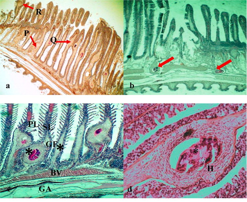
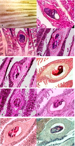
ig. 3 Histopathological changes caused by C. formosanus on the gills of koi carp. (a) Gill of koi carp: Note the encysted metacercariae in the base (P), middle (Q) and the apical portion (R) of the gill filament (×10). (b) Metacercariae encysted on the gill arch (arrow) with hyperplastic tissue (×40). (c) Distortion of gill lamellae due to fusion of the hyperplastic tissues (*) surrounding the parasites which located in adjacent the lamellae (×40): Primary Lamellae (PL), Secondary Lamellae (SL), Blood Vessel (BV), Gill Filament (GF) and Gill Arch (GA). (d) Parasite (*) is obvious in the center of chondrodysplasia (H) (×100, H& E).
ig. 4 The sequence of chronological lesions caused by metacercaria of C. formosanus in the gills of C. carpio. (a) Cercariae on the gill filaments after 2 h of infection (×40, H&E). (b) After 15 h of infection. (c) After 24 h of infection. (d) After 48 h of infection. (e) After 72 h of infection. (f) After 4 days of infection. (g) After 5 days of infection (h) After 7 days of infection. (i) After 10 days of infection. (j) After 21 days of infection (from (b) to (j) – magnification power: X 100, H&E).
ig. 5 (a) Hyperplastic cysts in the gill filament without the parasite(×40, H&E). (b) Higher magnification of Fig. a (×100, H&E).
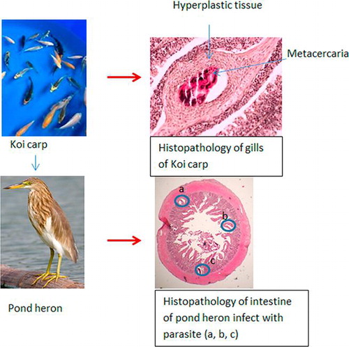
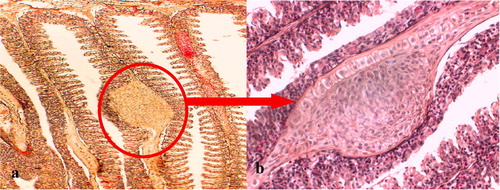
ig. 6 Invasion of C. formosanus into the superficial and deep layers of the duodenum of A. ralloides (×40, H&E).
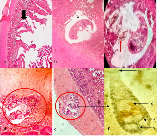
ig. 7 Invasion of C. formosanus into the intestine of A. ralloides. (a) Worm found in between the fused villus (×40, H&E). (b) C. formosanus in the sub mucosae (*) destroying the intestinal glands ( × 400, H&E). (c) Fluke with released eggs (arrow) (×400, H&E). (d) Parasite Invading the tunica muscularis (×400, H&E). (e) Invasion of C. formosanus into the deeper layer of the tunica muscularis of the intestine (×100, H&E). (f) Adult fluke of C. formosanus found in the small intestine of A. ralloides (p: Oral sucker, q: Eggs, r: Testes, s: Excretory bladder, ×100).