Figures & data
Figure 1 (A) Growth curve of B. burgdorferi strain B31 in vitro. (B) Representative images of the log phase (3-day culture) and stationary phase of B. burgdorferi B31 strain (7-day culture), observed with fluorescent microscopy using the SYBR Green I/PI stain (×400 magnification). The arrows indicate multiple morphological forms of B. burgdorferi in stationary phase.
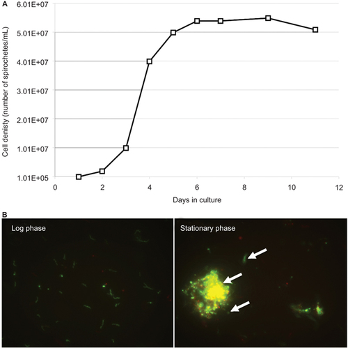
Figure 2 Susceptibility of log phase (3 days) and stationary-phase (7 days) B. burgdorferi to 50 µM drugs after a 5-day treatment. The percentages of residual live cells were determined using the SYBR Green I/PI assay.

Table 1 Activity of top 27 active hits with better activity than the current Lyme disease antibiotics against stationary-phase B. burgdorferi persisters Footnotea
Table 2 Comparison of the MIC values and anti-persister activity of selected antibiotics against B. burgdorferi
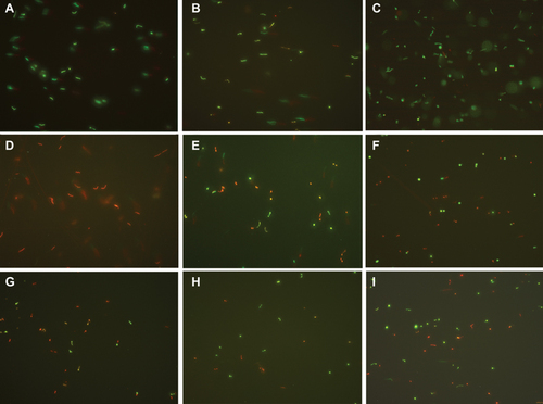
Figure 3 Representative images of stationary-phase B. burgdorferi strain B31 treated with different antibiotics (50 µM) followed by staining in the SYBR Green I/PI assay (×400 magnification). (A) Drug-free control, (B) Doxycycline, (C) Amoxicillin, (D) Daptomycin, (E) Cefoperazone, (F) Clofazimine, (G) Carbomycin, (H) Cefotiam, and (I) Tetracycline.