Figures & data
Figure 1 Microscopic evidence of babesiosis in a patient from the China–Myanmar border area. Giemsa stained thick blood smears (A) and thin blood smears (B and C) obtained on the first day of hospitalization for patient show an intraerythrocytic trophozoite (thin arrow). The lacking of hemozoin deposits distinguishes Babesia spp. from Plasmodium spp. The tetrad (thick arrow) is pathognomonic of small Babesia spp. Original magnification, ×1000.
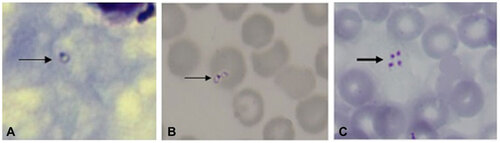
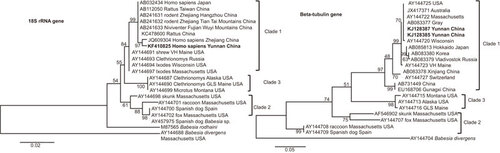
Figure 2 Phylogenetic trees of the SSU rRNA and beta-tubulin gene sequences for the B. microti isolates obtained from the two patients in the China–Myanmar border area. Phylogenetic analysis produced by the neighbour-joining method using MEGA version 5.1 software. SSU rRNA and beta-tubulin gene sequences of our case study samples were denoted as Yunnan China in bold face. B. divergens and B. rodhaini were set as the outgroups. Scale bar indicates nucleotide substitutions per site.