Figures & data
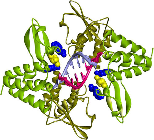
Figure 1 Structure of dsRNA C-terminal binding domain of EBOV VP35. Binding to dsRNA and IFN inhibition is dependent on a VP35 complex in which two VP35 molecules are paired in binding to a single RNA strand. Illustrated are the crystallographic coordinates of a truncated VP35 (residues 215–340) complex with an 8-bp dsRNA. Each dsRNA strand is depicted as red or blue-grey with similarly colored bases to indicate that binding of VP35 is nucleotide sequence independent. Three positively charged residues of VP35 are critical for binding to the phosphodiester dsRNA backbone, which results in inhibition of the initiation of innate immune responses including IFN induction. Arginines 312 and 339 on each binding homodimer (mint green) are represented as blue CPK van der Waals radii, with lysine 319 shown as a yellow CPK van der Waals radius. The second monomer (brown) of each pair does not make direct contact with dsRNA, although hydrogen bonds exist between the homodimers. Helical structures are represented as coils (α-helices) or flat bands (β-sheets). The VP35 binding affinity for dsRNA is size-dependent (2.8 nM for 500-bp dsRNA versus 3.2 nM for 50-bp dsRNA).Citation6 The illustration is based on PDB ID: 3L25 coordinatesCitation7 using Accelrys version 3.5 software. CPK, Corey–Pauling–Koltun.